No More Nitro

The truth is sometimes I enjoy a good medication shortage.  It forces us to come off of autopilot and think through the goals of therapy and how we can achieve them now that a commonly-used medication is unavailable.  Think about how much fun you all had using ketamine when we had a benzo shortage!  Shortages force us to get comfortable with unfamiliar medications, and look at you all now!  I’d be surprised to hear that one of you hasn’t used ketamine in some fashion.  Groovy stuff!

Shortages also create the opportunity to revisit the evidence behind certain therapies in order to reserve the medication for the most appropriate patients.  Who knows, you might uncover something that changes your practice.  Let’s take a look at what some smart people have to say about IV nitroglycerin use in acute decompensated heart failure:

Heart Failure Society of America 2010 Guidelines

Intravenous vasodilators (nitroprusside, nitroglycerin, or nesiritide) may be considered in patients with ADHF who have persistent severe HF despite aggressive treatment with diuretics and standard oral therapies.  

Nitroprusside (Strength of Evidence = B)  

Nitroglycerin, Nesiritide (Strength of Evidence = C) 

Intravenous vasodilators (nitroglycerin or nitroprusside) and diuretics are recommended for rapid symptom relief in patients with acute pulmonary edema or severe hypertension. (Strength of Evidence = C)    

European Heart Failure 2012 Guidelines

An i.v. infusion of a nitrate should be considered in patients with pulmonary congestion/edema and a systolic blood pressure >110 mmHg, who do not have severe mitral or aortic stenosis, to reduce pulmonary capillary wedge pressure and systemic vascular resistance. Nitrates may also relieve dyspnea and congestion (Class IIb, Level of Evidence B).   

ACCF/AHA Heart Failure 2013 Guidelines

Intravenous nitroglycerin, nitroprusside, or nesiritide may be considered an adjuvant to diuretic therapy for stable patients with HF (Class IIb, Level of Evidence A).    

I’m seeing a lot of nesiritide and nitroprusside talk along with this nitroglycerin (and shocked to see an A rating on evidence from ACCF/AHA).  When we look at the references cited for grouping nitroglycerin in with the other two medications, we find that there is VERY sparse data for nitro.  It’s all nesiritide and nitroprusside data. For nitro, there’s one nonrandomized, open-label, feasibility study of high-dose bolus nitro in 29 patients, and a 1998 Israeli study of 104 patients that is riddled with protocol violations.  There is one other trial comparing nesitiride vs. nitro called the VMAC study.  Spoiler alert: nesiritide outcompeted nitroglycerin.

If you are scared of the potential adverse events of nesiritide (elevated serum creatinine, questionable trend towards mortality in a meta analysis) or nitroprusside (thiocyanate toxicity…SUPER rare), know that nitroglycerin has potential harm as well.  In fact, in that VMAC study, more patients in the nitro group experienced adverse effects than with nesiritide.  Also, 7-day and 6-month mortalities were similar in the nitro and nesiritide group for VMAC.  Thirty-day readmission was similar with both as well.

Nitro tolerance and resistance are also big problems not seen with the other two.  Initially a benefit may be seen but over the course of hours, there may be no further improvement with subsequent increases in dose.

Additionally, keep in mind that many of these studies occurred before BiPAP became more popular for managing heart failure patients (we thought it led to a higher mortality, then it seemed to reduce mortality, now it may seem as though it leads to a higher mortality again…that’s for you all to determine).  I only point this out to show there has been a change in management for these patients since most of this data was published.

To further bash nitro, in a relatively recent review article on nitroglycerin for acute decompensated heart failure, the authors flatly conclude that there is currently not enough evidence to recommend nitro as a standard treatment.     

My bottom line for judicious nitro administration:

Nitro has NEVER been shown to have a mortality benefit in ADHF, weight risk vs benefit     

Patients presenting with concomitant ischemia take precedence  

Couple it with a good dose of diuretic

Reserve it for patients requiring BiPAP or intubation  

Reserve it for patients with an SBP of at least 120    

Start at a LOW dose and titrate up every 5-10 minutes to max of 200 mcg/min      

Avoid hypotension! Hypotensive events = mortality in these patients. BiPAP will drop their pressure. Give them a minute to have those million molecules of oxygen bombard their lungs. Their work of breathing will decrease, their endogenous catecholamine surge will subside, their pressure will go down.

Give the nurse clear instructions for titrations and explain the goals of therapy  

STAY IN THE ROOM FOR A HOT MINUTE   

Titrate the drip with the nurse for a while to see how the patient is responding to therapy

Get weird with it and request some nitroprusside or nesiritide!  Increase your drug repertoire, increase your knowledge, increase your comfort level  


Feel free to approach me with any questions or concerns about the medication management of heart failure patients.  I can also get you my references if you want your mind blown.    

Forget what ya’ heard.  Nitro may not be the saving grace you may think it is.

CK here- you may be thinking, What? Nesiritide kills people and NTG is awesome, everyone knows that. just look at http://crashingpatient.com/medical-surgical/cardiology/heart-failure-acute-pulmonary-edema.htm/

Or at least nesiritide doesn't work [see  http://www.thepharmacyconnection.com/cardiology/2011/07/07/is-this-nesiritides-last-call-for-acute-heart-failure-ascend-hf-published/ or read the ASCEND-HF trial in NEJM 2011].

Neal, however, has a well reasoned reply:

NTG:  We do not use it as they do in these studies.  I’ve never seen high-dose boluses of NTG in our ER.  I’m not sure how that blog crashingpatient gets away with using strong wording like “very effective” when referencing a trial of 9 patients.  I have been noticing a regurgitation of Scott Weingart anecdotes that are only based on a couple of his patients and his expert opinion…the same level of evidence he jokingly calls “GOBSAT (Good ole boys sitting around a table).”  
These studies have incredibly low numbers, most are non-randomized, convenience sampling, open-label, violate their pre-specified protocols, and don’t reflect current practice (i.e. non-invasive ventilation).
How is no one bringing any of this up?  There is clear bias towards utilizing NTG.  My argument in the post is that this bias is at least partially due to unfamiliarity with other medications.
Nesiritide:  I didn’t mean for it to seem that nesiritide is a clear winner over NTG.  I agree the data is not great.
Nitroprusside: Where are the criticisms for this medication in the setting of ADHF?  I personally believe that more people are silent on this because there is some data advocating the use but got overshadowed by ease of NTG, thus falling to the wayside.  This is a great candidate to replace NTG if there was no more NTG.
The whole point of this is to have critical responses.  The culture is to give NTG.  The levels of recommendation were put in there to show that evidence is sparse and most are in context of diuretic administration (people love to bash diuretics, but fail to admit there is just as big a void of data for nitrates).  I personally don’t care how much NTG gets used when we have it, whether the patient is a good candidate or not (just as long as they don’t get symptomatically hypotensive).  What I advocate is the responsible use of a dwindling product.

The use of sodium bicarbonate in patients with Severe Lactic Acidosis

The use of Sodium Bicarbonate in emergency Situations

The utility of intravenous sodium bicarbonate in severe metabolic acidosis is to say the least controversial. Yet each time we are presented with a patient with severe acidosis it is almost irresistible to give the patient an ampule of sodium bicarbonate to “improve myocardial performance”, after all our endogenous catecholamines work better under more physiologic conditions, right? 

I made this review to help delineate the problems of treating acidosis with sodium bicarbonate in the patients with acute metabolic (specifically lactic) acidosis. This is not meant for patients with renal tubular acidosis or patients who have bicarbonate loss, in which bicarbonate therapy is generally accepted (usually oral replacement). Also this is not for toxicologic emergencies, or for use in treating hyponatremia.

When considering bicarbonate replacement we must first consider the following questions:

  •  What are the complications of metabolic acidosis?
  • Can bicarbonate raise the pH?
  • Are there any beneficial effects of sodium bicarbonate?
  • What are the deleterious effects of sodium bicarbonate therapy?


What are the untoward effects of metabolic acidosis?

Before you read on think out loud what in your mind are the detrimental effects of metabolic acidosis and we are not talking prognostically here. 

-Reduced myocardial performance and decreased sensitivity of the myocardium to catecholamines, probably due to a reduction in the number of beta-receptors on the myocardial cell surface. In multiple isolated animal studies this has not been confirmed. This is because serum acidosis does not always translate into worsened intracellular acidosis. Also acidosis improves oxygen unloading into metabolically active cells. 

-Arrythmogenic especially at a pH < 7.2. This makes resuscitation of individuals in ventricular fibrillation more difficult, and why bicarbonate therapy was adopted during cardiac arrest. Oh the rise and fall of medications during ACLS! 

On the other hand we know that most patients tolerate respiratory acidosis fairly well. Everyday we place patients on ventilator settings to optimize their PaO2, and allow the PCO2 to rise without significant untoward hemodynamic effects (permissive hypercapnia). At a pH < 7.0 this is less well characterized, since so few people allow patients to develop such acidemia. 

Also MANY patients in DKA are extremely acidemic and do quite well  (how many times have you seen a patient with DKA with a pH < 7.2 who walked out of the hospital in 2 days?)

Conclusion: Therefore the assumption that a low pH in it of itself is a precursor to hemodynamic collapse and requires treatment is false.  This does not mean you should not recognize the acidemia as a potentially poor prognostic sign, and a marker of severe disease.  


Can Bicarbonate raise the pH?

Bicarbonate itself is not the only determinant of blood pH, as everyone knows. 

If you follow the physiochemical method of acid base disorders the pH is determined by subtracting strong cations (Na, K, Ca and Mg) from strong anions (Cl- and SO4). The SID increases due to bicarbonate administration because of an increase in sodium (bicarbonate is not a strong ion at all). 

However, bicarbonate drives Le Chatelier’s principle of chemical equilibrium: (HCO3 + H+ <==> H20 + CO2) 

 which will drive CO2 production. Therefore if ventilation is fixed, or MAXED as in a patient with a Minute ventilation of 20L, excess CO2 production cannot be eliminated and a rise in CO2 negates any potentially beneficial effects of the bicarbonate itself.

IF that gave you a migraine let me say it in another way. 

Sodium bicarb does increase the serum pH but it also increases CO2 production (think about ETCO2 jumping during a code when giving bicarb if you do this) and if you cannot blow off the CO2 with increased ventilation it has no net effect on the serum pH.

*Another potentially MORE IMPORTANT problem with following the serum pH is that different compartments have different pH’s (i.e; inner mitochondrial membrane, blood brain barrier etc) and CO2 readily crosses many of these membranes whereas bicarbonate itself does not. Therefore are we making the central veins for sampling more alkalemic at the cost of worsening intracellular and cerebral acidemia? 

Multiple studies have demonstrated that sodium bicarbonate will worsen acidosis in the brain and CSF (LP sampling and spectroscopy MRI). Several animal studies have shown that intracellular pH drops in RBC’s, muscle, liver and lymphocytes. 

Conclusion: Yes bicarbonate can increase the serum pH, but its effects on intracellular pH are unknown, but likely worsen acidosis. 


What beneficial effects does sodium bicarb have, if any?

Few studies have addressed specifically if there is any beneficial effect of sodium bicarbonate in vivo. No RCT’s have shown improvements in mortality, there has however been conflicting results on its effect on arterial pH, and several studies have demonstrated untoward hemodynamic effects.  

In animals sodium bicarbonate was indistinguishable from saline bolus on hemodynamics (CO, MAP, and CVP)

In two human studies in patients with lactic acidosis, sodium bicarbonate raised serum pH and bicarb, but did not improve hemodynamics or catecholamine responsiveness (ScVO2, MAP, wedge pressure, CO unchanged when compared to saline). 

Bicarbonate is an excellent volume expander, since it is like giving hypertonic saline, so potentially it could be use for resuscitation. Also bicarbonate can be given as a bolus for patients with severe hyponatremia with neurologic findings (about the equivalent of 100 cc of 3% saline in one 50 meq ampule.) 

Conclusion: Bicarbonate infusion has not definitively shown beneficial effects in patient or surrogate outcomes like hemodynamics or amount of vasoactives being infused . In fact most studies suggest bicarb has no net effects other than volume expansion in the hemodynamically unstable.


The final question remains what are the potentially deleterious effects of administration of sodium bicarbonate?

The first time I brought up the utility of sodium bicarbonate in the cardiac ICU, I was met with 

“ DO you know how many times I have regretted giving sodium bicarbonate? That would be 0”.

Essentially clinicians believe it is a benign cheap medication, which has few adverse effects. Oh but so wrong you can be:

Case:

55 y/o M s/p crush injury who had recently come off CVVH and was now making more urine whose bicarbonate had continued to be low (15). He was on the vent, but because of his non-gap metabolic acidosis he was breathing significantly above the vent settings to drive his PCO2 down to compensate for this acidemia.  This was making weaning difficult, as the attending did not want to extubate a patient with a RR in the 30’s. 

The surgical intern on overnight thought he would order a sodium bicarbonate drip in order to improve his serum bicarbonate and pH to make him “ready” for extubation. 

The resident ordered a 50 meq/hr drip at 8pm and at 5am labs were sent. When I arrived for rounds a critical sodium value was 168 (increased from 132). His chloride was also 130 so we thought this was an error, unfortunately a stat ABG had a pH of 7.67 and a HCO3 of 52 from the excessive endogenous bicarbonate that was given. The resident had MEANT to order a bicarb drip (3 amps in a liter of D5W or sterile water) which has a total of 150 mEq in 1L, instead he ordered 50 mEq/hr! 

The patient was less arousable after stopping sedation and continuous EEG found the patient to be in non-convulsive status (which alkalemia predisposes you to). It was UGLY. 

Detrimental effects of bicarb:

  • Increased serum sodium
  • Increased serum osmolality
  • Hypocalcemia can be profound
  • May worsen lactic acidosis
  • Overshoot correction and metabolic alkalosis


Conclusion: Bicarbonate will raise the serum pH when used in patients with severe acidosis. However, it is more likely that bicarbonate infusion worsens intracellular pH and does not provide any beneficial hemodynamic effects. Therefore in patients with LACTIC acidosis (even at extremes of pH < 7.2) it is difficult to find any reason to recommend sodium bicarbonate therapy. 

You are better off attempting to divert more neurons to solving why they are so acidemic. So the next time your nephrologist calls and screams why they aren’t on a bicarb drip you can kindly explain that there literature the use of bicarbonate is at BEST controversial. 


-Dave


Treatment of Acute Appendicitis-Antibiotics versus Surgery

Many thanks to Sheila and Ron Bonaguro for hosting, and to Catherine, Kasia, Katie B, Erin, Jen B and Lindsay for their excellent discussion of the articles.

As background, there have been numerous low quality studies published dating back to 1959 raising the question of treating acute appendicitis with antibiotics rather than surgery.   This journal club analyzed a meta-analysis of 4 studies (Varadhan), a Cochrane systemic review of 5 studies (Wilms) and probably the best original study on the topic  (Vons).  Surgery is not without risk, and the goal of JC was to evaluate if available literature supports antibiotics as a safe, curative strategy for acute appendicitis.  NB, all literature discussed dealt only with adult patients.

Overall Bottom Line:  The strategy of primary antibiotic treatment appears safe, and may obviate the need for surgery in some patients with acute appendicitis, but at least a quarter of patients will still go on to need surgery within the next year.  Consensus in the room was that for healthy adults, surgery is the preferred treatment.  One contrarian (EK) prefers antibiotics.  For a subset of patients who are poor surgical risks, it would be reasonable to engage the patient/family in patient centered decision making regarding the option for antibiotic treatment.

Future:  there are 2 ongoing prospective trials of antibiotics for appendicitis (APPAC and NOTA), which will likely provide higher quality data down the road.


1.  Varadhan KK, Neal KR, Lobo DN:  Safety and efficacy of antibiotics compared with appendicectomy for treatment of uncomplicated acute appendicitis : meta-analysis of randomised controlled trials. BMJ  2012; 2156:1–15. 

This meta-analysis illustrates the “garbage in-garbage out” phenomenon of meta-analyses.  This should be an extremely high level methodology for evaluating literature on a topic, but if the trials evaluated are low quality, then the meta-analysis conclusions will be very limited.  Authors evaluated 4 RCTs, with a total of 900 patients (approx half who received antibiotics and half surgery), and concluded that antibiotics are both “effective and safe as primary treatment for patients with uncomplicated acute appendicitis.”  While the antibiotic strategy appeared safe with regards to complications such as perforation, peritonitis and wound infection, 37% of patients receiving antibiotics ultimately required surgery for appendicitis within the following year...not so effective.  One issue with the studies was the variable (or no) imaging used to diagnose appendicitis, calling into question the accuracy of the diagnosis of appendicitis.  If some patients labeled appendicitis didn’t have appendicitis, antibiotics would end up looking very effective.  Overall a number of comments were made about the poor quality of the included studies, including the significant cross-over between groups in one study,  enrollment only of males in another, and significant followup issues.  And, as Chintan pointed out, our established intervention, surgery, is safe and effective (post-op complication rate 9-11%, 10 year adhesion/obstruction rate 3%); we’re not looking for an alternative to a dangerous or inadequate intervention. 


2.  Wilms IM, de Hoog DE, de Visser DC, Janzing HM. Appendectomy versus antibiotic treatment for acute appendicitis. Cochrane Database Systemic Review. 2011;11(11).

So, a plug for Cochrane reviews.  They are usually very high quality, and include a “plain language summary” which provides a quick and dirty conclusion about the topic.  In this review of 5 RCTs/quasi RCTs on antibiotics for acute appendicitis, they performed a non-inferiority analysis to evaluate if antibiotics are as effective as surgery in acute appendicitis.  It included some of the same trials as the meta-analysis.  Total of 901 patients; 73% of patients treated with antibiotics compared to 97% of patients receiving surgery were cured within 2 weeks without major complications at one year.  Study quality was low to moderate, with variability in diagnostic evaluation and use of prophylactic antibiotics.  Due to the pre-defined non-inferiority margin and overlap of confidence intervals for the outcome measures between the 2 groups, their conclusion is that surgery remains the standard treatment for acute appendicitis.  Antibiotics might be ethically studied as a treatment arm in a RCT, or considered in specific patients where surgery is contraindicated.

Side note about non inferiority analysis....these are often performed in drug company trials as the need is only to show that a new treatment is as effective or at least only marginally less effective than the old treatment.   A rather arbitrary margin of non-inferiority is defined by the investigators.  Burden of proof is less than in superiority trials.  For more details, please see Dr. Burns.


3.  Vons C, Barry C, Maitre S, Pautrat K, Leconte M, Costaglioli B, et al. Amoxicillin plus clavulanic acid versus appendicectomy for treatment of acute uncomplicated appendicitis: an open-label, non-inferiority, randomised controlled trial. Lancet 2011; 377(9777):1573–9.

This non-inferiority RCT enrolled 243 adults with acute uncomplicated appendicitis at 6 university hospitals and randomized them to surgery or augmentin x 8 days.  The study was included in both the meta-analysis and the Cochrane review, and is probably the best single trial out there.  All patients received CT scan for diagnosis.  Primary outcome was post-intervention peritonitis within 30 days of treatment, and occurred in 8% of the antibiotic group versus 2% of the surgery group.  In the antibiotic group, 26% of patients had recurrence of appendicitis and required appendectomy between 1 month and 1 year.  Authors concluded that antibiotics were not non-inferior to appendectomy for acute appendicitis, and that surgery remains the gold standard treatment for acute uncomplicated appendicitis.  There were no significant differences between the 2 groups for postintervention complications.  Interestingly, presence of fecalith was predictive both of complicated appendicitis in surgery patients as well as antibiotic failure in the medical group (fecalith = bad).  Plenty of issues with this study as well-no discussion of timing of surgery, all surgery patients also received pre-op antibiotics, and 18% of patients diagnosed with “uncomplicated appendicitis” by CT in the surgery group had complicated appendicitis with peritonitis identified in the OR.  One year followup was missing for 10% of patients.

56 YO MALE WITH SOB

HPI: 56 yo male with small cell lung cancer on chemotherapy (3 days ago, first tx) presents to the ED with SOB over the past day. No chest pain, LE edema, orthopnea. No fevers, chills or hemoptysis. +Chronic dry cough.  He also complains of generalized weakness with ntermittent nausea, vomiting and diarrhea since chemotx. No abdominal pain. No urinary changes.

Physical Exam:

VS: HR 124, BP 102/69, Sat 89% on RA, RR 30, T 36.5 R

GEN: Alert, mild-mod distress, thin

HEENT: MM dry

CV: tachy, regular

PULM: Course bilaterally with wheezing. Tachypneic. No rales

ABD: Soft, NT

EXT: No LE edema

INTERVENTION #1: O2, BIPAP, nebs, 2 L NS bolus

EKG: Sinus Tach. No STE or right heart strain

CXR:

CXR1 1.jpg

“There are extensive new airspace opacities at the lung bases and left upper lobe . . .”

INTERVENTION #2: Vanc, zoysn and azithromycin for HAP. Unsure of WBC at this point. HR and RR improving with IV fluids, Bipap and O2. BP is stable. Still somewhat tachypneic.

LABS:

Na

132

WBC

57.2

UA =

unremarkable

K

2.8

Hb

11.2

BNP

314

Cl

90

Hct

33.7

Trop

0.37

Bicarb

19

Plt

498

Flu

Neg

BUN

52

PMN

95% = 56

C diff

Neg

Creat

2.31

Bands

3

L.A.

3.0

Ca

5.6

Glc

184

***So why is the calcium so low? Kessen, who is sitting next to me and fresh off of his oral boards beat down by Kerwin (ONC emergencies) says, “tumor lysis syndrome”.

-But what about the potassium? Shouldn’t it be higher?

-So we add-on a few labs:

Uric Acid

9.2 (3.5-7.2)

Phos

7.8 (2.4-4.7)

Mg

0.8 (1.6-2.4)

-Well shit, it is tumor lysis syndrome (TLS), right?

INTERVENTION #3:|

-Consult oncology

-Replace K, Mg, Ca.

-IVF hydration

-After a discussion with Neil (pharmD), we order Rasburicase for the elevated uric acid (Apparently it’s very expensive ~$20,000)

-Oncology agrees with assessment and plan. States that TLS is associated with small cell lung tumor

-Admitted to MICU

HOSPITAL COURSE:

-Repeat Uric Acid is 3.6 after 6 hours

-other electrolyte abnormalities are more difficult to correct, but do improve by day 3

-Pts respiratory distress worsens and he is intubated the 1st day in the ICU

-he become neutropenic and pancyotpenic on day 3-4 (~7 days after chemotx). Possible DIC

-Pt dies on day 7

 ---------------------------------------------------------------------------------------------------------------------

TUMOR LYSIS SYNDROME (TLS): from uptodate.

Massive tumor cell lysis with the release of large amounts of potassium, phosphate, and nucleic acids into the systemic circulation. Catabolism of the nucleic acids to uric acid leads to hyperuricemia.

TLS most often occurs after the initiation of cytotoxic therapy in patients with high-grade lymphomas and acute lymphoblastic leukemia. However, TLS can occur spontaneously and with other tumor types that have a high proliferative rate, large tumor burden, or high sensitivity to cytotoxic therapy.

-Hyperuricemia: a consequence of the catabolism of purine nucleic acids to hypoxanthine and xanthine and then to uric acid via the enzyme xanthine oxidase. Results in the precipitation of uric acid in the renal tubules leading to acute kidney injury

-Hyperphosphotemia: rapid tumor breakdown often leads to hyperphosphatemia which can cause secondary hypocalcemia. This can lead to AKI by calcium phosphate deposition in the renal tubules and also to cardiac arrhythmias.

DEFINITION:

Cairo-Bishop definition of laboratory tumor lysis syndrome

Element

Value

Uric acid

≥ 8 mg/dL

Potassium

≥6.0 mmol/L (or 6 mEq/L)

Phosphorus

≥ 6.5 mg/dL for children or 4.5 mg/dL for adults

Calcium

≤  7 mg/dL

- Two or more laboratory changes within 3 days before or 7 days after cytotoxic therapy

TREATMENT:

-Correct electrolytes and monitor q 4-6hrs

-Hyperkalemia: typical treatment, including dialysis if severe

-Hyperuricemia: IVF and rasburicase (0.2 mg/kg, repeat as necessary). Rasburicase is urate oxidase (uricase), which catalyzes oxidation of uric acid to the much more water-soluble compound allantoin. Can also do Allopurinol if rasburicase is not available.

-Hypocalemia: Treat only if symptomatic. Correct elevated phoshate first if possible

-Hyperphoshatemia: Oral binding agents (not very effective)

-Kidney injury: IVF rehydration, treat elevated uric acid. May need dialysis.

GCBH2...57 yo F...psych complaint

57 yo F brought in by brother-in-law for purported suicide attempt. He tells me she's done things in the past, but nothing serious, and that he believes she is just trying to get attention. Pt is somnolent but arousable, I do my quick standard psych H&P, patient reports that she drank 10 ounces of windshield wiper fluid approximately 12 hours ago with the intent of killing herself. She is mildly tachypneic and diaphoretic, so the first the thing we do is order an ABG:

pH 7.06, HCO3 6, and anion gap of 32

ELEVATED ANION GAP ACIDOSIS!!

Remember MUDPILES?

M-methanol

U-uremia

D-DKA

P-propylene glycol

I-infection/isoniazid/iron tox/isopropyl alcohol

L-lactic acidosis

E-ethylene glycol

S-salycylism

Re-evaluation of patient showed decreasing GCS and increased WOB, so we moved her to CC room and intubated her. Patient was started on fomepizole drip (I wanted to started pouring whiskey down her OG tube but Erik wouldn't let me) for presumed toxic alcohol toxicity, vascular surgery was called to place catheter for emergency HD,  and nephrology was emergently consulted.

Toxic alcohol levels showed an initial methanol level of 145 mg/dL; no ethylene glycol or isopropyl alcohol detected.

Nephrology recommended adding bicarb drip, and within a couple hours hemodialysis had begun. After 3 hrs of HD, methanol levels were down to 68, pH increased to 7.50, and the anion gap was closing. Pt then went to ICU.

She underwent a total of 12 hours of HD. On day 2 of hospitalization, methanol level was <25, so fomepizole and HD were dc'ed. Pt was shortly therafter extubated, and transferred the next day to psych floor. Interestingly, she never developed any opthalmologic complications.

One final footnote: Before she crunked and got tubed, she was trying to remember the brand of wiper fluid she'd guzzled. All she could remember that it had the word "Blast" in it. After some internet research, I read that there was indeed a company based out of Minnesota that made a wiper fluid called "PowerBlast." Interestingly, they were currently being sued by another windshield wiper producer for "watering down" their product, ie, using less methanol, to save on manufacturing cost. I found it interesting that perhaps by working to improve their bottom line, the company had inadvertently made their product less toxic, and as such perhaps prevented this particular patient's demise (although who knows the M&M that resulted from spinouts out on icy roads by drivers unable to see out of frozen, wiper-fluid-encrusted windshields).

What's the level?

TOXICOLOGY CASE OF THE MONTH

FEBRUARY 2014

55yoM h/o seizure disorder, Hepatitis C, HTN, DM, CKD, meningioma s/p resection and hepatic encephalopathy presents from assisted living for mental status changes. It is his second visit in the last 48 hours for similar symptoms. He has been difficult to arouse and was noted to have an unsteady gait. He denies fall or intentional ingestion. He is unsure whether he has had any recent medication changes. He complains of generalized weakness and unsteadiness.

Medications: Albuterol, Atorvastatin, Clonidine, Gabapentin, Hydralazine, Hydroxyzine, Insulin, Lactulose, Nifedipine, Phenytoin, Terazosin

Physical exam:

VS 97.2 142/84 69 18 100% RA

General: Awake, alert and oriented x3 though unsure of purpose of hospitalization, no acute distress, cooperative

HEENT: PERRL - 4mm to 3mm b/l, EOMI, + b/l horizontal nystagmus

Neck: supple no asymmetry

CV: RRR no m/r/g

Lungs: CTA b/l

Abdomen: + BS, soft, nontender,nondistended

Extremities: WWP, 2+ pulses in b/l radial, PT, DP, no clonus, no asterixis

Neuro: CN II-XII grossly intact, 5/5 strength in b/l UE and LE, sensation intact b/l UE and LE, wide-based ataxic gait with b/l dysmetria on finger to nose

Skin: warm, dry, no diaphoresis, no rash or lesions

1. What is this patient’s differential diagnosis?

  • Non-tox causes: hepatic encephalopathy, infection, incranial mass/hemorrhage, CVA
  • Toxicology differential:
    • Most likely antiepileptic given presentation - Phenytoin or Gabapentin
    • Hydroxyzine can cause somnolence but would expect anticholinergic presentation (elevated temperature and HR, dry skin and mucous membranes, delirium, urinary retention, etc)
    • Clonidine can cause somnolence but would expect different toxidrome (opioid-like - MS depression with respiratory depression, + VS abnormalities - hypotension & bradycardia)

2. What is the differential diagnosis of nystagmus in toxicology?

  • Many drugs and toxins cause nystagmus - usually horizontal. Here are a few examples:
  • Anti-epileptics
    • Barbiturates
    • Carbamazepine
    • Phenytoin
    • Gabapentin
    • Ethanol
    • Scorpion Envenomation
    • Isoniazid
    • PCP (horizontal, vertical, rotatory)

Horizontal Nystagmus

Labs:

Initial presentation:

  • 140/4.4/109/25/44/2.21/214
  • AST 30 ALT 41 Bili 0.2 Albumin 2.5
  • Phenytoin 14 mcg/mL (ref range 10-20)
  • Ammonia 25

Second presentation:

  • 136/4.8/107/22/49/2.0/200
  • AST 41 ALT 31 Bili 0.1 Albumin 2.5
  • Phenytoin 20 mcg/mL (ref range 10-20)
  • Ammonia 17

 (Creatinine is at baseline)

CT head with no acute change

3. How do these labs change your differential diagnosis? What is the culprit agent?

  • The patient's signs and symptoms are consistent with Phenytoin toxicity
  • Phenytoin is >90% Protein-bound - level needs to be corrected for serum albumin
  • This patient is at risk - hypoalbuminemia due to liver disease

4. What are the clinical characteristics of phenytoin toxicity?

  • Mild to moderate: horizontal nystagmus, ataxia, dysarthria
    • Less common: nausea, vomiting, diplopia, hyperglycemia, agitation, irritability
  • Severe: stupor, coma, respiratory arrest
    • Rare seizures have been reported (though should always consider an alternative cause of seizures in a phenytoin-poisoned patient)
    • Death extremely rare
    • Gingival hyperplasia is indicative of chronic use - may clue physician into diagnosis
    • Reported cause of Stevens-Johnson Syndrome and Toxic Epidermal Necrolysis
Dilantin-induced-gingival-hyperplasia.JPG
  • Propylene glycol component in IV preparations can cause myocardial depression - hypotension, bradycardia
    • Cardiac toxicity does NOT occur with oral overdose or with fosphenytoin

5. What are the applicable pharmacokinetics?

  • Absorption can be slow and unpredictable and in acute ingestion peak effects may be delayed
  • 90% protein bound
  • Hepatic elimination – zero order kinetics near therapeutic range – half-life increases as levels rise

6. What is the formula to correct the serum phenytoin level?

  • Corrected Phenytoin = Measured phenytoin level/(0.2 x Albumin + 0.1)
  • Available on most medical calculators
  • This patient's correct levels are 23 and 33 respectively using this formula

7. How is phenytoin toxicity managed?

  • Hold offending medication
  • Most importantly: protect the patient from self-injury caused by ataxia
    • This may require the patient to be admitted to the hospital for fall prevention
      • For PO toxicity - do not need to be admitted to a tele floor (common misconception)
      • Can also d/c home with reliable family member if close follow up with neurologist or PMD can be arranged, if circumstances are appropriate
      • If seizures occur - consider alternative cause, treat with other anticonvulsants (benzodiazepines generally recommended for toxicology-related seizures)
      • If hypotension occurs with IV administration - stop infusion, administed IV fluids/vasopressors as necessary
      • No antidote, no role for dialysis. May consider activated charcoal if appropriate though most often toxicity is chronic.

Case Conclusion:

  • Send-out free phenytoin level was 4 ug/mL (ref range 1-2), % free 21.6% (ref range 8-14%)
  • Culprit medication was held and patient recovered clinically after several days
  • Was discharged home with a lower medication dose

Pearls to remember:

  • Phenytoin toxicity si/sx: ataxia, nystagmus, dysarthria
  • High protein binding - level should be corrected for serum albumin
  • Treatment is supportive - fall prevention, hold medication, dose adjustment
  • Cardiac monitoring not necessary for toxicity due to oral preparations

17 y/o with hip pain playing basketball

17 y/o male brought to PED2 by EMS after hurting his hip playing basketball. He was running down the court when his friend collided with hip, hitting friend's elbow on patient's bony pelvis. Patient said he couldn't walk afterwards, his dad seems to think he's being overly dramatic. No PMH. VSS. Exam shows tenderness on right side of bony pelvis without brusing or deformity. No other abnormalities on exam.

AP pelvis XRAY

ASIS.jpg

What's the diagnosis? Treatment?

OK, now that you've thought about it a while, I'm sure you've diagnoses an avulsion fracture of the anterior superior iliac spine or ASIS. These tend to occur in athletic adolescents who have a forceful contraction of the muscles that attach there (sartorious and tensor fascia lata). They fracture because the apophyses are forming in puberty. Other areas prone to avulsion fractures are ischial tuberosity, and AIIS (anterior inferior iliac spine). 

Xrays are often not obtained as the mechanism (sprinting, jumping) sounds like it wouldn't cause bony injury, and the injuries are misdiagnosed as a muscle strain. The good news is that the treatment is generally similar- rest with partial weight bearing on cructhes, pain control, followed by PT. If the bony fragments are more than 3 cm appart or healing is delayed, the patient is a candidate for surgical repair.

Treatment of Massive Pulmonary Embolsim

Treatment of Massive Pulmonary Embolism

CASE:

21-year-old male was admitted to saint elsewhere with severe shortness
of breath, epigastric discomfort and vomiting. He had recently been
admitted to outside hospital with pancreatitis for 10 days and was
discharged home with PMD f/u with improving idiopathic pancreatitis. 
When the patient arrived in the ED his vital signs were:

HR 140, Sat 92% on RA, BP 140/50

CC: epigastirc pain and vomiting

In the ED the patient was triaged to intermediate care had a chemistry
panel sent and a CT A/P ordered. 
His lipase came back at > 3000. The medicine team was consulted and
the patient was to be admitted to the floor pending a CT A/P. Image
shown below is the most superior image from the abdominal CT which
 happened to catch the main pulmonary arteries

The patient requires 4L NC to maintain his sats > 92% but is awake and mentating well and his BP was normal. His EKG is shown:

The ED felt the patient was hemodynamically stable to be admitted to the floor and medicine admitted with IR consults for possible
 thrombectomy and IA tPA due to large RV seen on CTA. About 5 am the next morning while in the angio-suite the patient codes during thrombectomy. The patient is PEA without an airway in IR.

If you are called to this code, what would you do? 
 

What was done:

A Pulmonary artery catheter was in place because of the attempted thrombectomy and the code team pushed 50mg of IA tPA 
(after about 10 minutes of me looking in the cath lab for the vial of tPA that was not in 2 mg aliquots). The patient has ROSC
 about 30 seconds later. Unfortunately his first ABG intubated on the
 vent was as follows:

6.79/90/85 on AC 30/500/100%/5 
 

What would be your next move, and why do you think the pH and CO2 are so high?

We decided to add inhaled nitric oxide (see below) with only mild improvements in the CO2 to 75, pH still 7.0 on 3 vasopressors. What would you do?

CV surgery was called for immediate ECMO, and the patient was cannulated in the angio suite. 

2 days later the patient was decannulated he did suffer from hemorrhagic conversion of his pancreatitis, and large RP hematomas and intra-abdominal hemorrhagic ascites, likely due to heparin and tPA administration.

However the patient was weaned off the ventilator, his pancreatitis improved and he was discharged home with no residual deficits on hospital day 21.  

SO what can you do in the ED to set the patient with massive PE up for success?

Disclaimer: I should mention this portion pertains to the specific subset of patients who have right ventricular failure and not simply pulmonary hypertension from a PE and are reasonably well compensated. These people have the findings I mention below.

Some tricks of the trade are as follows:

Although hypotension is almost always treated initially in the ED with IVF's if you suspect massive PE the cause of hypotension is RV failure/dilation and the treatment is RARELY IVF.

1. You will not have hypotension from acute massive PE without RV dilation

This is because the thin highly compliant RV whose afterload is the wimpy pulmonary arterial tree is very pressure sensitive. Rapid increases in pulmonary pressure result in massive increases in RV volume (it does not hypertrophy acutely). Since the right and left ventricles SHARE the septum the right ventricle balloons outward and impairs diastolic filling of the LV when pressure overloaded, hence the D shaped LV seen on parasternal short axis. This impairs the LV filling, reducing the mean arterial blood pressure and cardiac output.

Signs of RV failure are:

1. Distended neck veins/elevated CVP/ Dilated plethroic IVC without resp variation

2. Elevated transaminases (usually more chronic)

3. Hepatomegaly, lower ext edema

4. Kussmal's sign (sudden increase in CVP in spont breathing patients with inspiration)

Echo:

Dilated RV with wide-open tricuspid regurgitation (this is because of the stretch of the annulus of the valve NOT valvular dysfunction per se). Adding additional volume will only overload the RV even more and worsen TR. LV is usually hyperdynamic with small chamber size.

-To complicate matters the RV (which is usually a low pressure ventricle) gets coronary perfusion during both systole and diastole (since it is a low pressure ventricle). However as the RV pressure increases the coronary filling is markedly reduced during diastole, this creates RV ischemia. Ischemia results in stiffness of the ventricle making the RV more dysfunctional and so continues the downward spiral of right heart failure.

2. The treatment of significant RV failure:

-Reduce RV Preload

To reduce preload avoid giving volume in the ED and consider diuresis

Commonly diuretics are given if the CVP is significantly elevated because the normal geometry of the ventricles has been distorted due to an enlarged RV bowing the spetum towards the LV, diuresis will result in improved LV performance even with a decrease in intrasvascular volume.

-Reduce RV afterload:

Inhaled Nitric Oxidie or Inhaled Prostaglandins like Epoprostenol. Usually iNO is given starting at 20ppm, despite the fact that 90% of patients will have maximal response at 5ppm or less.

The problem with giving systemic pulmonary vasodilators in a hypotensive patient should be intuitive, the nice thing about giving inhaled pulmonary vasodilators is they only act where there is ventilation and perfusion. Nitric oxide is rapidly metabolized by local RBC's and thus has no systemic effects like hypotension.

iNO is not standard of care per se, but is a trick up your sleeve for that severely dyspneic patient with massive PE who you want to avoid intubation in. AM I saying you can give iNO non-invasively, absolutely. This is the nice thing about iNO is sometimes it will save you an intubation or at worst in can prevent the precipitous RV failure that will ensue after intubation.

-Maintain MAPS > 65

I know this is an obvious point, but it has to be stressed that even a minute of hypotension can throw a tenuous RV into the spiral of death mentioned. Sedatives will often result in rapid hemodynamic compromise if you are not careful.

Epinephrinebetween 0-50 nanos/kg/min or (0- 0.05mcg/kg/min) at lower doses epinephrine acts as an inotrope, and above that tends to act more as an alpha agonist. At low doses epi will likely not increase the pulmonary vascular resistance but at higher doses (> 50 nanos) it may. 

Dobutamine2-5 mcg/kg/min can augment RV function without increasing the PVR seen with epi and levophed, however it can cause systemic hypotension from beta 2 agonism and often will be given with another agent.

Vasopressin can augment the mean arterial pressure, and likely has NO effects on the PVR.

My unevidenced based practice is to start Dobutamine and Vasopressin (2 mcg/kg/min and 0.04 U/min) to keep MAPS > 70 and follow serial bedside echos.

3. If someone with a PE is dyspneic should I intubate sooner rather than later, they are eventually going to tire out right?

The problem with intubation in a patient with a PE is positive pressure ventilation increases the afterload via PEEP to the right ventricle. This may tip the RV over into florid RV failure.

There are also the hemodynamic perturbations with giving a sedative during induction, which can cause hypotension, again resulting in rapid hemodynamic compromise.

Initial paralysis will prevent spontaneous ventilation and may make it difficult to match a patient's own intrinsic minute ventilation (young pt breathing RR 40, and large TV of 1L with MiV of 40), try setting the vent to get anything reasonable in these patients afterwards is not ideal. If you have to use something use a shorter acting agent like succinylcholine. 

Nothing like someone telling you not to do something every sense in your body is telling you to do. However, if you are forced to intubate them, and you know they have a massive PE, consider thrombolysis up front and:

1. Start iNO, if this process isn't available where you work but iNO is available in house find a way to expedite the process in the ED now before you are really desperate. If you work in a hospital without iNO/CT surgery/IR consider establishing transfer to a facility with higher level of care. Worst thing you can do is admit a patient to an ICU where they won't be seen by an intensivist until the morning and there is minimal backup if the patient decompensates. Without a backup if the patient codes the game has been lost.

2. Have your vasopressors ready to go on a PUMP and push dose pressors in hand epi is the simplest in these situations.  I use epinephrine in 10mcg aliquots as a push until I have a MAP > 65 if they drop during intubation. Then I place and titrate a drip as needed.

3. Sometimes flipping into A-fib will result in rapid hemodynamic deterioration and may need cardioversion as the loss of the atrial kick may have significant hemodynamic effects.

4. Call your CV surgeon and IR attending and get them on board early. "I have a young 
patient with massive PE who is crumping in the ED, is this patient a candidate for surgical thrombectomy and
 if they crash, would you consider them an ECMO candidate?" 

Never be afraid to ask for help and if they are young and salvageable don’t accept NO for an answer. 

Eastvold Pearl #28 - Lithium Toxicity Masquerading as Wellen's Syndrome

Take-home point: Rule out lithium toxicity in all patients on lithium presenting with cardiac toxicity and/or EKG changes.

[1] The case: 48 yo F with history of bipolar disease and depression presents to ED for evaluation of substernal chest pressure radiating to neck with bilateral arm "numbness."  Mild shortness of breath and ROS only positive for mild gradual onset headache.  Symptoms x 7 hours.  Chest pain non-exertional, non-positional, and non-pleuritic.  No fever, focal neurologic deficits, mental status changes, or abdominal pain.  Chest pain 7/10 upon arrival, and improved with 3/10 with nitrate therapy but continued to have ongoing pain/symptoms.  Serial EKG revealed dynamic change, and concern for proximal LAD lesion and Wellen's syndrome was discussed with cardiology.  Cardiology performed a bedside echo which revealed anterior wall motion abnormality, and to a lesser degree inferior-lateral dyskinesia.  Patient was taken to cath lab for elective cath, with knowledge of an elevated troponin of 0.61 (normal < 0.03),  Cardiac cath revealed completely normal coronaries, and no LV aneurysm  Lithium level came back elevated at 2.3, with no history of acute ingestion.  Quick literature review (articles attached) revealed that lithium can mimic ischemic EKGs, especially in the anterior leads.  Further, chronic lithium toxicity can also cause myocarditis.  See EKGs below illustrating deep anterior T wave inversions and prolonged QTc, both of which resolved after lithium level decreased.

[2] Quick skinny on lithium toxicity and EKGs, attached is are 2 review articles as well.

  • Lithium causes toxicity through several mechanisms including competition with many electrolytes (sodium, potassium, calcium, and magnesium) and ion transporters, which can disrupt many cellular functions.  The cellular membrane of concern here is myocytes (and Purkinje fibers....and most people just closed this email!), and these disruptions manifest as EKG changes.   
  • Common EKG changes include [a] QTs prolongation, [b] nonspecific ST segment and T wave abnormalities, [c] T wave inversions (reference #1), [d] transient ST elevations mimicking acute myocardial infarction (reference #2), [e] Brugada changes with possibility of sudden cardiac death (reference #3), [f] SA nodal dysfunction and heart block, and [g] severe bradycardia.
  • ECG changes are more likely to occur with chronic lithium overdoses than acute overdoses
  • Treatment is beyond the scope of this review but some important things to consider are
    • Make sure you get lithium levels on all patients presenting with cardiac symptoms
    • Know the difference between acute vs chronic toxicity, read attached article
    • Know your criteria for hemodialysis
    • Avoid thiazides and ACE-I, as these will increase lithium levels
    • Avoid NSAIDS, can also worsen toxicity.  Often given for myocarditis, but do not given if you think this is lithium-induced myocarditis
    • Check TSH, can be contributing to lithium toxicity
    • Consult toxicology and nephrology early, especially if cardiology is going to manage primarily

Feedback always welcome (joshua.eastvold@gmail.com),

Also, if anyone is interested in publishing this let me know??

Josh 

References:

[1] Cases J. 2008 Sep 17;1(1):156. doi: 10.1186/1757-1626-1-156.

[2] J Med Toxicol. 2008 Sep;4(3):170-2.

[3] Circulation. 2010 Aug 10;122(6):e418-9

[4] Toxicol Rev. 2006;25(4):221-30.

[5] J Am Soc Nephrol. 1999 Mar;10(3):666-74

EKG #1 upon arrival to ED

wellens Li1.png

EKG #2 - concerning for anterior ischemic.  QTc 494.  

wellens Li2.png

EKG #3 - worsening QTc prolongation, still getting PO lithium, don't ask

wellens Li3.png

EKG #4 - Courtesy call to cardiology, relayed my thoughts regarding concern for chronic lithium toxicity

-- now after lithium level returned to 1.3, complete resolution of anterior T wave changes and normalization of QTc

wellens Li4.png

Intubating the Hypotensive Patient

Intubating the Patient with Hypotension

Before you intubate your next hypotensive patient, think twice and read below.

Positive pressure ventilation has multiple hemodynamic affects on your patient, mostly reduced venous return resulting in reduced preload, and increased right ventricular afterload. The combination of these two along with the systemic affects of your sedation for induction can result in rapid hemodynamic compromise in the already unstable patient.

Case:

58 y/o M with cough fevers and chills with body aches presents to the ED.

VS: 40.1, 135 (ST), sat 90% on NRB and he is breathing a real 30x’s/min, BP 85/55.

You determine the patient would benefit from endotracheal intubation, how do you proceed?

Step 1: Emergent or not?

The first question you always have to ask yourself is this going to be emergent, and do I have time. As ED doctors we are quick to jump to intubation when we know someone needs it, but the reality is most patients can be intubated urgently rather than emergently, while getting everything you need prior to induction. If they are agonal, severely hypoxic and cannot be BVM ventilated then godspeed.

But if you decide this is an urgent intubation, and have 5 minutes proceed

Step 2: What is my access?

The nurses should have 3 minutes to get you two working IV’s before you go to a humerus I/O. If you use the tibia be sure you use a pressure bag to augment the infusion rates, which usually are 65 ml/min vs. 165 ml/min with a pressure bag. In the Obese, sickle cell patient, or patient on hemodialysis this is the rapid IV of choice and results in fewer errors than placing a sterile semi-emergent CVC.

Why 3 minutes?

-Because while that is happening another nurse should be grabbing you two bags of crystalloid and a pressure bag ready to give to this patient.

-If you have a pharmD ask for a vasopressor drip on the pump ready to go, if they can make phenylephrine fastest or epinephrine take what you can get. Otherwise someone should be getting central pharmacy to make you drip, this is preferable to you making the drip yourself.

By now you should have two IV’s and one of the IV’s should be designated for induction medications. Have one bag of crystalloid running wide open on the IV that has been established as the line that will have the meds pushed through. This way we know if the IV is infiltrated (the bag isn’t running, or the skin around the IV has blown up) or if it just so happens that the BP cuff is on the same arm as the designated IV you realize before pushing the meds  (cause’ its just embarrassing trying to figure out why your patient isn’t paralyzed).

Now this is where I don’t expect most ED doctors to follow, but I would recommend this. Place an arterial line. You can argue there have been no studies that show that A-lines save lives and that it is a waste of time and not an ED skill, and you may be right. What I will argue is that’s true of chest x-ray’s, MRI’s and most innovative technologies and it is all about what you do with the information that makes the difference.

 The A-line gives you beat-to-beat information. There is no recycling of the blood pressure because the cuff couldn’t read and people start feeling for a pulse. CPR? Hit restart, that can’t be right! I know this has happened to all of us.

An US guided A-line doesn’t take much time to put in, once you are familiar with the technique. I believe it allows for a smoother more hemodynamically stable intubation. Are they hypertensive during laryngoscopy, (your unsecured aneurysms that have leaked but not ruptured!?) give him more propofol and take the blade out. Has my patient’s pressure just dropped? bolus him phenylephrine or epinephrine.

This also makes getting an ABG after intubation much easier than asking RT to do radial A-line sticks in hypotensive patients and prevents the dreaded; oh we couldn’t get an ABG so we didn’t do one.

Step 3: Induction Medications:

You can use whatever you want, just know they all cause hypotension. Ketamine seems to be en vogue, and is a good choice, but since we don’t have it in our omnicell in the ICU most of the ICU anesthesiologists here use an opiate like fentanyl first followed by a low dose of propofol; as in 10mg boluses up to 30mg until they are unresponsive to jaw thrust. I personally don’t think 20mg of etomidate is the best answer, but don’t have any science to back this up.  Use whatever you like and markedly reduce the dose of the sedative.

Step 4: You were ready for hypotension

It was going to happen, but now atleast you are ready for it. First continue your fluid bolus, which should now be on a pressure bag if the MAP starts to get a little soft.

Then if it is apparent that there is a loss of vasomotor tone, you should replace the lost endogenous catecholamines with temporary vasopressors. I suggest blousing aliquots of phenylephrine 100 mcg Q 1 minute unless the MAP has precipitously dropped, then start with 300mcg. Once you have stabilized their MAP see if you have to keep pushing phenylephrine, if you do they need a drip, if you don’t congratulations you have successfully intubated a sick hypotensive patient without killing them.

If you are slick with the echo probe and see the LV or RV is down consider epinephrine 10mcg boluses instead and an epi infusion if needed.

Step 5: How do I sedate my hypotensive patient?

Let me start off by saying I think there is enough good evidence to recommend avoidance of benzodiazepines completely in most ICU patients not seizing. BZDs result in increased delirium, prolonged ventilator days, and worse patient outcomes compared with alternatives. This is never truer than in the elderly.

With that said the first thing to address is pain. It’s uncomfortable to sit in a firm bed without moving for hours, while having a toothbrush shoved in the back of your throat and the IV monitor beeping over and over again. If you treat pain first you will have a marked reduction in the requirement for “sedation/amnesia”. I like a dilaudid push of 1mg after hemodynamics have settled out followed by a drip 0.5-2mg/hr.

The sedation choice for me is between low dose propofol (10-30 mcg/kg/min) and dexmedetomidine. I prefer dex, 0.1-0.7mcg/kg/hr and it is going generic this year. If people give you a hard time about choosing this “expensive medication” show them the data on benzodiazepines and hospital LOS, and ventilator days. If you use ketamine, as I know ACMC has, you are ahead of the curve.

When giving these meds you don’t need to SNOW the patient, just make them calm enough that they can be awoken with loud verbal stimuli (we use the RAAS scale, target -1 to -2).  Having them be comatose makes repeat exams with fluctuating mental status difficult to monitor.

This isn’t the sexy part of medicine but if you are in the business of saving lives and making your patients comfortable the nuances listed above is worth the extra effort.

The most important part of intubating the hypotensive patient is to be prepared for the rapid hemodynamic consequences that will follow. You shouldn’t sit after an intubation followed by PEA arrest wondering why that happened, it’s usually right in front of your face.

SIDE PEARL HYPOTENSION from obstructive shock:

If by chance you know the patient has a large pulmonary embolism as the cause of their distress you should be aware that intubation is likely to result in rapid hemodynamic deterioration. WHY? The RV is a low-pressure, high compliance chamber and rapid increases in afterload (PE) result in RV dilation. The RV dilation results in bowing of the interventricular septum to the left and causes reduced compliance of the LV, and the LV becomes more underfilled.  Since the RV is perfused during diastole and systole, any reduction in MAP (caused by LV dysfunction) will cause a stiffening of the RV again impeding forward flow. The now more dilated RV causes tricuspid annulus dilation and significant tricuspid regurgitation and again impedes forward flow. Intubation will exacerbate the RV failure by decreasing MAP, and increasing RV afterload especially if they become hypoxemic or hypercarbic as both increase pulmonary vascular resistance.

RV failure.png

Dave

"I think he took his gout medicine..."

JANUARY 2014

TOXICOLOGY CASE OF THE MONTH:

CASE:

58-year-old male presents to the hospital complaining of abdominal cramping, nausea, and vomiting.  He has a history of depression with suicide attempts in the past. He told his daughter he “may have taken some of his gout medication” earlier that day. His family says one of his bottles of medication, recently prescribed as needed for gout flares, is now empty. They are unsure how many pills were in the bottle.

XD141137.jpg

PMH: Gout, depression, hypertension

Meds: Hydrochlorothiazide, Colchicine, Allopurinol, Buproprion

All: NKDA

VS: 105/60 110 20 99% RA 36.6

Exam remarkable for tachypnea and dry mucous membranes. Lungs are clear. No focal abdominal tenderness though the patient does complain of cramping. Rectal exam with brown stool, guaiac positive.

Labs:

CBC: WBC 3.5, Hgb/Hct 11.3/34.1, Platelets 210

Chem: 132/3.2/95/14/45/1.2/110

LFTs wnl

Lactate 4.3

APAP/Salicylate/EtOH undetectable

Applicable case questions:

1. What medications are used to treat gout flares?

  • Colchicine
  • NSAIDs
  • Glucocorticoids (i.e. Prednisone)
  • Anakinra, an interleukin-1 antagonist, is used off-label
  • Note: Allopurinol is used for gout prevention, not in acute flares

2. What is the mechanism of action of colchicine? How is it prescribed?

  • Colchicine acts at a cellular level by arresting dividing cells during mitosis
  • It is used for treatment of gout and familial Mediterranean fever
  • It is also found in certain plants (Colchicum autumnale – meadow saffron)
  • It is rapidly absorbed into body tissues
  • FDA-approved therapeutic dose: 1.2mg orally, followed by 0.6mg after 1 hour
  • Should not be prescribed long term
colchicine.jpg

3. Why should I be worried about it?

  • Significant toxicity with no antidote and no effective means of elimination
  • One case series described the following:
    • 0.5 mg/kg: diarrhea and vomiting
    • 0.5-0.8 mg/kg: bone marrow aplasia and 10% mortality
    • >0.8 mg/kg: uniformly resulted in death
    • Fatalities have been reported with ingestions as little as 7mg (though some have survived with ingestions >60mg)

4. What are the clinical effects of an acute colchicine overdose?

  • Many organ systems effected from hours to days after exposure
  • 2-12 hours after presentation: GI symptoms
    • Nausea, vomiting, abdominal pain, diarrhea (often bloody)
    • Diarrhea: common side effect of therapeutic use
    • Progresses to shock: fluid loss and depressed cardiac contractility
    • Delirium, seizures, and coma
    • Lactic acidosis due to shock and inhibition of cellular metabolism
    • Myocardial injury
    • Rhabdomyolysis
    • DIC
    • Renal failure
    • Bone marrow suppression, alopecia, polyneuropathy (late effects)
    • Death after 8-36 hours by respiratory failure, intractable shock, and cardiac arrhythmias

5. How is colchicine toxicity diagnosed?

  • Clinical diagnosis
  • History of gout and familial Mediaterranean fever
  • Severe gastroenteritis closely followed by leukocytosis, shock, rhabdomyolysis, and acute renal failure followed by leukopenia and thrombocytopenia and general decompensation
  • Levels not readily available (forensic use primarily)
  • Other useful labs: CBC, CMP, CPK, UA, Troponin, EKG, Lactate

6. What is the management?

  • Aggressive supportive care and shock treatment
  • Anticipate respiratory and cardiac collapse
  • Highly fatal overdose with no antidote and no effective means of removal (cannot use hemodialysis)
    • Colchicine-specific antibodies (Fab fragments) tested in France, not commercially available
    • Rifampin is a CYP3A4 induced – may enhance elimination in theory but no good data
  • One of the only times in toxicology where aggressive decontamination is highly recommended
    • Charcoal, gastric lavage, whole bowel irrigation are considerations
    • Undergoes enterohepatic recirculation: may need multi dose activated charcoal
    • Some recommend intubation for aggressive decontamination
    • If survive initial insult may need treatment for bone marrow depression

7. What are the acute toxicities of other agents used to treat acute gout flares?

  • NSAIDs
    • Pharmacologic and toxicologic effects occur via COX inhibition
    • Usually asymptomatic or GI upset
    • May develop significant CNS depression, seizures, renal failure, acidosis, hepatic dysfunction with large ingestions
    • Usual treatment is IV fluids and H2 blockers
    • Hemodialysis not effective
    • Glucocorticoids
      • Small ingestions: usually GI upset
      • Anxiety, agitation
      • Fluid retention
      • Majority of toxic effects due to chronic use (i.e. Cushing’s syndrome)

CASE CLOSURE:

  • The patient developed hypovolemic shock
  • Intubated and aggressive decontamination with charcoal via NG
  • Ultimately death with v-fib arrest

TAKE-HOME PEARLS:

  • Colchicine is a highly toxic medication used to treat gout and familial Mediterranean fever
  • Presentation: gastroenteritis followed by shock, renal failure, and cardiorespiratory collapse, late bone marrow suppression
  • Aggressive supportive care and decontamination recommended
Gout-Cartoon_Beer-Consumption_2_Bar_CLR.jpg

Eastvold Pearl #27 - Superficial thrombophlebitis in the lower extremity

Hey guys,

Been a while, and have 2 more coming.  

Superficial thrombophlebitis (ST), aka superficial vein thrombosis

--

Thrombosis of superficial veins has long been considered benign, and deemed a separate entity from venous thromboemolism (VTE)

-- However, multiple studies illustrate a significant association with VTE (DVT and PE).

-- When patient with ST (diagnosed clinically, no ultrasound) are thoroughly evaluated, the degree and the extent of clot are underestimated 75% of the time.  Further, such patients are found to have co-existent DVT or PE 25% of the time and/or rapidly progress to DVT 10% of the time.  

---- Teaching point: get ultrasounds on all clinical superficial thrombophlebitis

-- The risk factors for ST and VTE are the same, and many argue that ST should be treated the same as VTE.  

-- In general,

anticoagulate (as you would for DVT) the patient if they have known clotting risk factors, greater than 5 cm of clot, or clot < 5 cm within the sapheno-femoral or sapheno-popliteal junction.

-- Another way to think about it is that a superficial vein thrombosis is a manifestation of a systemic clotting cascade gone awry.  To even further simplify things, seems pretty pathologic to have any blood vessel clot; ahh hello, you are clotting off blood vessels.

Below is my treatment algorithm on ST in the lower extremity.

Feedback always welcome.

Josh

References

http://www.ncbi.nlm.nih.gov/pmc/articles/PMC3278049/#

    CHEST 2012 guidelines

Eur J Vasc Endovasc Surg 2005; 29:10-17. 

J Vasc Surg 2003; 37:834-8. 

Thromb Haemost 2001 ;86:452-63.

SVT.jpg

* Full anticoagulation with [a] lovenox 1 mg/kg BID (or 1.5 mg/kg QD) for at least 5 days and until coumadin is therapeutic for at least 24 hours. [b] Xarelto or rivaroxaban15 mg BID for the first 3 weeks, then 20mg daily for at least 3 months.  If lifelong anticoagulation planned, would recommend lovenox/coumadin.  2nd reoccurrence consider lifelong anticoagulation but refer to CHEST guidelines for specific permutations. A study by Schulman found a second DVT indicated a need for lifelong anticoagulation and it didn’t matter whether the DVT was provoked or unprovoked.  However, provoking factors vary in their risk of recurrence; surgery patients have the lowest risk of recurrence.  Patients with unprovoked or idiopathic thrombosis or multiple recurrences with lower risk provoking factors will get long‐term anticoagulation. 

** History of DVT or PE, active malignancy (recommend solely lovenox, d/w oncologist), hypercoagulable state.  Other risk factors that warrant consideration of systemic anticoagulation include male gender without varicose veins, history of superficial thrombophlebitis, recent surgery, involvement of saphenous vein, and severe symptoms.

*** Lots of variability in dosing. 

  -- Lovenox: [a] 40 mg daily SC x 4 weeks per CHEST guidelines, [b] 1 mg/kg BID SC x 10 days then 1 mg/kg QD x 20 days per Belgian Society on Thrombosis and Haemostasis. 

  -- Fondaparinux 2.5 mg SC QD x 30-45 days

  -- Xarelto – current trial comparing 10 mg PO QD vsfondaparinux.  Xarelto approved for DVT.  Would consider using if 1st time on anticoagulation given PO form and no need for INRs, but would discuss with PCP before committing to lifelong use of Xarelto.

****

Deep veins = superficial femoral vein, common femoral vein, deep femoral vein, popliteal vein, anterior tibial vein, posterior tibial vein, peroneal vein.

Superficial veins = Greater saphenous vein, small saphenous vein

Left Ventricular Assist Devices

Left Ventricular Assist Devices:

I'm on call in Cardiothoracic ICU, so if figured I would take a minute to see if I can drop some pearls regarding the management of left ventricular assist devices, since its fresh in my mind.

First what is a left ventricular assist device (LVAD from here on out):

Essentially an LVAD is a device that is used in patients with severe left ventricular dysfunction. If you have pre-operative biventricular dysfunction you are not a candidate for an LVAD, and instead may be a candidate for heart transplant (pending your age usually young like < 65 y/o, good renal and liver function). Because patients with NYHA class IV heart failure become significantly malnourished and develop end organ dysfunction a lot more patients are going to total artificial hearts (we use syncardia) as a bridge to transplant (BTT) and to keep the patients mobile with good end-organ perfusion until a suitable donor is found (which can take a significant amount of time.) These will become more popular over the next few years and are a whole different animal to manage.

There are multiple types of LVADs although most have gone to continuous flow centrifugal pumps, most frequently used is the HEARTMATE II, although some places have started using the Heartware. The older pulsatile pumps have fallen out of favor and were outperformed by the continuous flow pumps with a 58% survival at 2 years vs 24% in the continuous and pulsatile flows respectively.

Slaughter MS, Rogers JG, Milano CA, et al. Advanced heart failure treated with continuous-flow left ventricular assist device. N Engl J Med 2009;361:2241-2251

The LVAD is essentially connected to the left ventricular apex, and to the ascending aorta. See picture:

LVAD image.jpg

The patient then has a driveline that is connected to the rotor (located subdiaphragmatic), which exits the patient’s abdomen and connects to their battery pack.

Therefore since the LVAD is a continuous flow device, which unloads the left ventricle continuously, patients may or may not have a pulse.

The pulse is created by opening and closing of the aortic valve as it is directly related to their own intrinsic contractility. If you place an A-line you may see it be mostly flat with a MAP, hopefully, > 65. If a patient starts to regain cardiac function (myocarditis patients) then it is possible to have a strong pulse even with an LVAD. It is important to know that the aortic valve may not open in some patients, and that the patient’s hemodynamics don’t depend on it opening!

OK now that some of the basics are out of the way lets give some of the meat, what are the numbers on the device?

Pump speed (RPMs)

-This is how fast the rotor is moving usually somewhere between 8600-10,000 rpms. This is not picked arbitrarily. The RPMSs essentially is a major determinant of your cardiac output. SO some might say why not just go up higher on the RPMs if the patient is hypotensive? The answer is because the VAD is essentially a vacuum and if the cavity of the LV is too empty it may pull on the free wall or septum of the LV. These are called "suction events" which can precipitate dysrhythmias (have you ever put a guide wire in too deep and irritated the RV with frequent PVCs, try pulling on it with a vacuum).

The normal geometry of the LV should have the inflow cannula (going to rotor) facing the mitral valve without rightward or leftward shift of the septum or free wall.

If the patient becomes hypovolemic the LVAD may start precipitating suction events, because the RPMs are set too high for the relative amount of volume and pull on the septum. The VAD accommodates for these events by temporarily going down on the RPM's to try and reset itself.

PEARL: if the RPM's are changing on its own their it is likely a SUCTION EVENT, think hypovolemia or dysrhythmia.

RECAP: A ramp study (a study where someone “ramps up the RPMs” under echocardiography is required to change the RPMs because the geometry of the LV must be watched real time and therefore the RPMs should rarely be changed in the emergency department without an expereicned VAD provider doing so under echo guiadance.

The POWER (watts)

This is essentially how much power is required for the device to continue running at the set RPM's. There is no one number as it varies between patients and trending this number is usually the most helpful, the patients generally know where they have been running.

Why is the power important? The power and the RPMs are the only numbers on the VAD devices that are NOT GUESSTIMATES. All other numbers are derived from these two values.

If the power is increased it is because more work is required to continue turning the rotor at a fixed rate.

What could possibly cause the rotor to require more power to maintain the same set RPMs? Well the VAD is a synthetic device that is prone to clotting (about 1% incidence). If the INR is low, and you don't hear that nice hum of the VAD on auscultation, and instead hear a trapped weasel (I swear this is the closest thing I can imagine this as) maybe its a clot on the rotor. The machine is still trying to turn at constant RPMs but is pushing against a clot so more power is required.

Other causes of increased power are an increase in speed (RPMs) increase in volume status or an increased afterload.

What if the power is low, well you can imagine this means that the rotor is seeing less volume, this could be caused by hypovolemia or a CLOT either pre or post- cannula. A clot in the rotor will cause an increase in power, where as an LV thrombus or a thrombus on the outflow cannula will cause a drop in power.

Pump Flow (L/min)

The flow is a guesstimate of CO. Let me repeat that the FLOW is a guesstimate of the cardiac output. The flow depends upon volume status, LV contractility, the speed, RV function and afterload.

The heartmate II will alarm at low flow states < 2.5L/min and will show --- and flows that are above physiologic limits for a set RPMs just present as +++ on the LVAD.

Why is this important, well if there is a clot on the rotor, the machine has increased power, the same RPMs and the CALCULATED flow will be high even though the real flow will be far less. The machine knows this and gives you a clue to this by presenting +++ as a flow estimate since it is outside the physiologic range.

As mentioned before this is a flow estimate and is not analgous to cardiac output. 

What is the Pulsatility Index? (dimensionless value)

Now things get really hairy. The flow is a calculated flow based on power and RPMs. And the PI is calculated from flow (max flow- min flow/avg flow over 15 seconds). So its calculated from a calculated number!

What it’s supposed to represent is the intrinsic contractilty of the LV. As the LV increases its contractility the flow will theoretically increase as more blood goes through the LVAD.

Joe Hsu my mentor puts it this way:

As the LV contracts, the flow through the pump increases due to an increased pressure at the pump inlet approximating the pressure at the outlet cannula (aortic pressure). During cardiac diastole, this inlet pressure drops, while the outlet pressure remains high (increased pressure difference) and, consequently, flow decreases.  Therefore, pulse index is directly proportional to amount of LV contractility (increased due to preload, inotropic support, and myocardial recovery) and inversely proportional to the assistance provided by the pump.

So what you need to take from this is the PI estimates the amount of LV function, and as the patient becomes hypovolemic often times the PI will drop with their MAPs.

What are common LVAD complications?

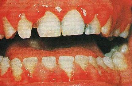
1. Acute Anemia/Bleeding:

You have all seen this and is the most common reason patients with LVADs present to the ED, especially with GI bleeding. The question always comes up can you reverse the INR, and the answer is of course yes. If the patient has a significant bleed you should consider reversing it. They can bridge to heparin or integrilin in the ICU if need be when the patient is more stable. We do not start heparin for 5 days in fresh post-ops and a lot of times mid-ICU course they go unanticoagulated for days, and people don't clot off.

Hemolysis is another cause, which can cause significant anemia especially with a pump thrombus. LDH, haptoglobin, bilirubin as well as cell-free plasma hemoglobin can aid if you are very suspicious.

Can I give too much blood too quickly if they are end stage HF? Yes and NO the LV, sucks but that’s why they got the LVAD and as long as the RV is working please give the blood rapidly. If you notice RV failure (significant JVD, hepatomegaly, lower ext edema, assuming you don’t have a CVC for a CVP, which the CVP is not completely dead in this circumstance) then I would urge caution. Which brings us to

2. RV dysfunction:

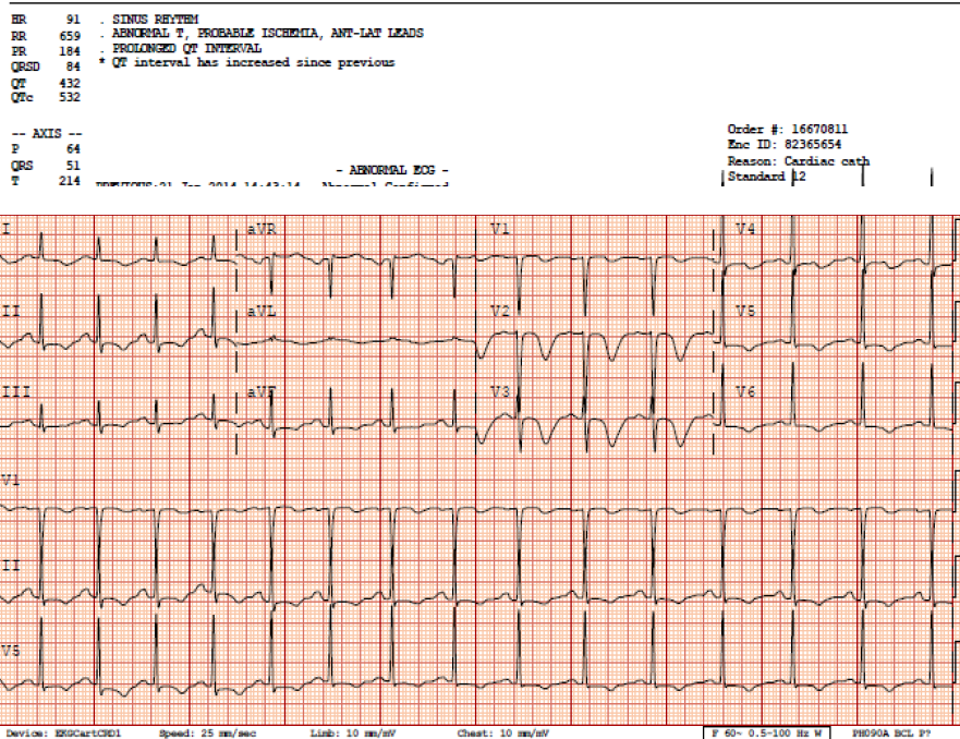
This is a complication in about 20% of post-op patients and sometimes they have to crash onto an RVAD. This is badness. If the patient is not hypovolemic, and they are hypotensive and have signs of RV dysfunction you need a stat echo or do a bedside. Get an EKG are they ischemic, are they sub therapeutic in INR, did they throw a PE, is the LV so big its causing the septum to bow into the RV? do your best to figure out why is the RV failing.

Remember these were sick hearts to begin with, the LV gets fixed with an LVAD and then the RV sees all this additional flow it wasn't seeing before, and now a sluggish but getting by RV can become sick. If you have RV dysfunction you have got problems, and need a surgeons help quick. Thankfully this won't be common as most of these occur quickly after surgery.

How can you manage RV dysfunction, well a lot of times milrinone will aid in reducing pulmonary vascular resistance (PVR), and increases RV contractility. If you need to intubate do so cautiously as the PEEP from mechanical ventilation will worsen RV dysfunction and you should minimize hypoxia and hypercapnia as these two entities will worsen PVR.

If you suspect RV dysfunction get a VAD specialist in the ED immediately.

3. Infections:

These can be ugly, depending on where they are. These patients get blood cultures if they are febrile because they have a device, and will need long-term abx if infected. Replacing an LVAD is a morbid procedure and not one people are likely to do if they can avoid it.

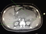
Be a good doc and examine the abdomen and the site where the driveline comes out since it can be your source if its related to the LVAD (not a viral syndrome). These should get a CT scan to evaluate for an abscess if you don't find another otherwise obvious source (skin/ua/chest), and plain CT C/A/P usually will suffice. We usually start BROAD spectrum abx vanco and zosyn, and because of the device if the patient is in septic shock consider up front adding anti-fungal coverage with an echinocandin (micafungin, caspofungin or anadilafungin).

4. Tachydysrrthmias:

Ventricular arrhythmias

Why did they get the LVAD in the first place, ischemic cardiomyopathy is more likely to have irritable myocardium, but NICM is certainly entitled. These don't go away because they have an LVAD, and VTACH or VFIB can precipitate suction events as the RV may not be able to coordinate an adequate contraction which reduces LV preload. These dysrhythmias can be managed with cardioversion, which is not problematic, but can cause myocardial stunning afterwards. If they are stable VTACH consider amiodarone/procainimide or lidocaine and most of the patients have their own ICD's from their prior CM so make them comfortable if possible. If one of the first two doesn't work we usually add lidocaine as a bolus.

AFIB with RVR

Afib will also be problematic reducing RV function and therefore LV preload most surgeons here opt for rhythm control with amiodarone bolus and drip since they are anti-coagulated already with INRs 2-2.5.

5.Cardiac Arrest

Can I do chest compressions? Sure you can, but remember the LVAD is taking care of perfusion to the body so solving why its not doing that should be a priority. PE, hemorrhagic shock, RV failure, tamponade, hypovolemia etc…

The risks of doing chest compressions are dislodging the inflow and outflow cannula which if that occurs will likely result in massive exsanguination. So if you have to do it, you have to do it but take a second and try and figure out why the patient has coded. Otherwise continue with ACLS as per usual.

6.Aortic Insufficiency:

This adversely affects the pumps function by causing rapid LV filling and high pump flow as the outflow cannula is valveless and merely follows the path of least resistance (low pressure LV vs. higher pressure systemic vasculature).

Patients can present with left heart failure, pulmonary edema and hypoxemia. Treatment for mild AI is usually afterload reducing agents and diuretics, but for mod-severe AI may require a new valve or over-sewing completely of the original aortic valve in an effort to reduce the regurgitant jet.

BELOW is a TABLE of common complications and treatment options, its good reference in-case you need to look it up on a shift its courtesy of Joe Hsu, Critical Care at Stanford.

OK that’s a lot of information but it will get you started. Please e-mail me with questions and I’ll post-em on the site. If you have had a particularly challenging LVAD case let me know and if I don’t know the answer I have the mentors who will.

Talk soon!

-Dave

Table 1: Diagnosis and Management of Adverse Events and Complications

Complication

Clinical features

LVAD Parameters

Diagnosis

Management strategies

Diagnostic Studies

CT scan findings (7)

Inflow Cannula Complications                                 

Kinking of inflow cannula

é Cardiac pulse pressure, Hypotension

ê Flow, power,

Variable flow

Coagulation parameters (INR 1.5-2.5), CT scan

--Kinking of inflow cannula

--Cannula malposition (Figure 6)

--Surgical consultation

--Anticoagulation

--Maintain adequate volume status

Thrombus in cannula

é Cardiac pulse pressure

Acute anemia (due to hemolysis), Hypotension

ê Flow, power

Evaluate for hemolysis: é cell-free plasma Hgb, indirect bilirubin, LDH; ê Haptoglobin

CT scan

--Low attenuation lesion in inflow/outflow cannula

--Surgical consultation

--Anticoagulation

Outflow Cannula Complications

Tearing of aortic anastomosis

Hypotension, Hemorrhage

No specific change

Serial Hgb, TEE, CT scan

--Extravasation of contrast material at anastomosis

-- Emergent surgical consultation

--Can occur over time

Kinking of outflow

Cannula

é Cardiac pulse pressure, Hypotension

ê Flow, power

Variable flow (positional)

Coagulation parameters (INR 1.5-2.5), CT scan

--Kinking of outflow cannula

--Disruption of cannula patency

--Surgical consultation

--Anticoagulation

Hemodynamic Complications

Arrhythmia

± Hemodynamic instability, Symptoms of ê perfusion

ê Flow, power

ECG, Electrolyte panel, TTE/TEE (to assess LV geometry, fluid status and cannula position)

None

--Control arrhythmia, defibrillation

--Evaluate for suction events-VT

--Watch for RVF

--External chest compression-onlyif in extremis

Right

Ventricular Failure

Hypotension, é CVP, PVR

ê Flow, power

Suction events

TTE, TEE, CT scan

CVC ± PAC

--Intraventricular septum bowed leftward,

--RV dilated,

--Dilation of IVC

--RV contractility: inotropes (Milrinone)

--Decrease PVR: avoid hypercapnia, hypoxemia, pulmonary vasodilators (iNO)

--Avoid overfilling

--Control arrhythmias

Pericardial tamponade

é CVP

Hypotension,

Preload dependent

ê Flow, power

Suction events

TTE, TEE (pericardial effusion may not be visualized on TTE), CT scan

--Dilation of IVC,

--Compression of right ventricle or atrium, flattening of heart border

--Pericardial effusion

--Pericardiocentesis

Aortic valve insufficiency

Decompensated heart failure, êSystemic perfusion, Cardiogenic shock

é High flow

TTE ramp study to evaluate speed, ECG-gated CT scan

--Presence of valvular thickening,

--Aortic valve visualized on ECG-gated CT scan

--May develop over time

--Consult cardiology and cardiac surgery

--Diuretics, afterload reduction

-- AI may improve with decreased pump speed

Coagulation-related complications        

Thrombus on Rotor

Acute anemia (due to hemolysis)

é Cardiac pulse pressure

+++” Flow,

éPower (>10-12 W)—hrs to days

Evaluate for hemolysis: é cell-free plasma Hgb, indirect bilirubin, LDH; ê Haptoglobin

CT scan

--Low attenuation lesion near inflow

--Surgical consultation

--Anticoagulation

Hemorrhage

Primarily GI also: Epistaxis, Hematuria, Mediastinal, Thoracic

ICH

ê Flow, power

Suction events

TTE, TEE, EGD, serial CBC, coagulation parameters, CT scan

Evaluate for acquired vWD: vWF antigen, vWF activity, factor VIII activity

Evaluate for hemolysis: é cell-free plasma Hgb, indirect bilirubin, LDH; ê Haptoglobin

--Evidence of bleed (e.g., ICH on CT head)

--PRBC and pro-coagulant factors as indicated

--Avoid excessive transfusions in:

    -Bridge to transplant

    -History of RVF

--?vWF replacement

Infectious Complications

LVAD-specific (41)

  • ·  Pump/cannula infections
  • ·  Pocket infections
  • ·  Percutaneous driveline infections

LVAD-related (41)

  • ·  Endocarditis
  • ·  Bacteremia
  • ·  Mediastinitis

Fever, Chills, Hypotension 2/2 sepsis,

Sequelae of embolic events

Variable effect on LVAD

CBC with differential, lactate

Exit site Wound Cx  (+ Fungal Cx): if risk factors

TEE, if TTE negative

Blood Cx, if + CVC obtain “Time to positivity”Cx

Imaging: Ultrasound, CT (c/a/p)

--Gas or fluid collection around pump components, percutaneous lead

--Figure 4, pocket infection

--Figure 5, percutaneous lead infection

--Broad spectrum antimicrobials for nosocomial pathogens

± Fungal coverage based on risk factors

--EGDT

--Surgical consultation for incision and drainage, debridement, device exchange

Abbreviations: CVC: Central venous catheter, CVP: Central venous pressure,  Cx: Culture, EGDT: Early goal directed therapy, GI: Gastrointestinal, Hgb: Hemoglobin, ICH: Intracranial hemorrhage, LDH: Lactate dehydrogenase, PAC: Pulmonary artery catheter, PVR: Pulmonary vascular resistance, RV: Right ventricle, RVF: Right ventricular failure, TEE: Transesophageal echocardiogram, TTE: Transthoracic echocardiogram, vWD: von Willebrand disease vWF: von Willebrand factor, W: Watts

Everything is toxic: the dose determines the (rat) poison.

DECEMBER 2013

TOXICOLOGY CASE OF THE MONTH:

CASE:

63yoF presents to the Emergency Department for her second time in a week complaining of epistaxis. On the first visit, she had packing placed. Bleeding was attributed to dry environment and changing weather. She was discharged home with Cephalexin. However, she returned to the ED 2 days later complaining not only of persistent epistaxis, but of hematuria and rectal bleeding. On further questioning, she admits to taking a “handful” of d-CON daily over the past 2 weeks as a suicide attempt.

PMH: Depression, HTN, no hematalogic disorders

Meds: Prozac, Amlodipine; no blood-thinning medications

All: NKDA

VS: 101/59125 16 99% RA 36.6

Exam remarkable for persistent epistaxis from L nare without a clear site of bleeding. Rectal exam notable for gross positive blood. No skin lesions (petechiae, purpura, ecchymosis).

Labs:

CBC: WBC 9.5, Hgb/Hct 9.2/27.9, Platelets 210

Chem: 132/4.2/95/26/55/0.8/110

LFTs wnl

INR: >15

Acetaminophen, Salicylate, EtOH undetectable

Applicable case questions:

  1. What is the Toxicology differential diagnosis of bleeding?
  2. What is d-CON?
  3. Why should I be worried about it?
  4. What is the toxic dose?
  5. What are the clinical effects? What labs should I order?
  6. What is the most appropriate management of rodenticide ingestions?
d-con

Answers:

1. What is the Toxicology differential diagnosis of bleeding?

  • Note: this differential dose not include other causes that should be considered such as bleeding disorders, hepatic failure, DIC, ITP, TTP
  • A lot of pharmacology but an important review – we see patients on may of these
  • Warfarins and suprawarfarins (i.e. rodenticide)
  • Direct thrombin inhibitors (ie Dabigatran/Pradaxa)
  • Factor Xa inhibitors (ie Rivaroxaban (Xarelto)
  • Anti-platelet agents
    • COX inhibitors (Aspirin)
    • ADP receptor inhibitors (Clopidogrel/Plavix, Prasugrel/Effient, Ticagrelor/Brilinta, Ticlopidine/Ticlid)
    • Glycoprotein IIB/IIA inhibitors: mostly IV use (Abciximab/Reopro, Eptifibatide/Integrilin, Tirofiban/Aggrastat)
    • Adenosine reuptake inhibitors (Dipyridamole/Persantine)
    • PDE inhibitors (Cilostazol/Pletal)
  • Rattlesnake envenomation
  • Arsenic toxicity (may have pancytopenia 1-2 weeks after acute ingestion, usually more leukopenia and anemia)

2. What is d-CON?

  • Anticoagulant rodenticide – contains long-acting suprawarfarins
    • Brodifacoum is most common: this is what is in d-CON brand
    • Others: Diphacinone, bromadiolone, chlorophacinone, difenacoum, pindone, valone
    • Interesting fact: warfarin initially used for rat poison, however rats and mice became resistant

3. Why should I be worried about it?

  • Like warfarin, suprawarfarins inhibit conversion of vitamin K to its active form: necessary to synthesize coagulation factors II, VII, IX, X, proteins C & S
  • The duration of anticoagulation from a single dose of warfarin is usually 2 to 5 days
  • Suprawarfarins may produce significant anticoagulation for weeks to months after a single ingestion

4. What is the toxic dose?

  • Warfarin
    • Single small dose (10-20mg) will not cause serious toxicity
    • Chronic or repeated ingestion (i.e. 2mg/day) can cause significant anticoagulation
  • Suprawarfarins:
    • Potential to have prolonged effects even after a single small ingestion
    • Large study of accidental suprawarfarin ingestion in children: no serious cases of ingestions less than 1 box
      • Ingels M, Lai C, et al. A prospective study of acute, unintentional, pediatric superwarfarin ingestions managed without decontamination.. Ann Emerg Med 2002;40(1):73-8.

5. What are the clinical effects? What labs should I order?

  • Excessive anticoagulation
    • Warfarin: anticoagulation effects within 15 hours, lasts up to 5 days
      • Suprawarfarin: Anticoagulant effects usually delayed up to 2 days and may persist weeks to months
      • CBC, PT/INR, Type & Screen (if bleeding),
        • Toxicologists will order Brodifacoum levels to determine duration of therapy
        • If you ordered warfarin-dependent clotting factors, these will be decreased
  • PTT, fibrinogen, liver enzymes, D dimer may help evaluatuation of other causes of bleeding if cause is unknown
  • Acetaminophen, Salicylate levels – rule out coingestants

6. What is the appropriate management of d-CON ingestions?

  • Most ingestions: accidental, by children
  • Poison center recommends: send child to the hospital if they ingest greater than a box of d-CON or other rodenticide, otherwise can be observed at home
    • Parents can observe for bleeding clinically
    • Poison center recommends that these patients can have a follow up at PMD office – a normal PT/INR 48 hours after exposure rules out significant ingestion (Studies suggest that if patients have no clinically evident bleeding that this measure may be unnecessary)
  • If patient comes to the ED just after this ingestion:
    • Activated charcoal
      • Appropriate conditions (recent exposure, normal mental status, not vomiting)
      • Not indicated if patient ingests less than 1 box
      • Labs will not give any information about toxicity but may establish baseline if there is concern for any underlying hematologic disorders
      • Do not give prophylactic Vitamin K (see below)
  • Significant bleeding
    • Treat shock – pRBC transfusion
    • Vitamin K
      • Give if evidence of clinically significant anticoagulation
      • Do not give prophylactically for any ingestion: if this is the case, then the 48 hour PT/INR cannot be used to determine severity -  will need at least 5 days of monitoring
      • Be aware: patients with significant bleeding due to suprawarfarin toxicity need weeks to months of treatment with Vitamin K – poison center can help admitting physicians with dosing but large doses may be required (up to 800mg per day!)
      • FFP for non-life threatening bleeding
      • PCC’s for life-threatening bleeding if available, otherwise FFP
      • Management can be difficult if patient requires long-term anticoagulation (ie prosthetic valve) – can consider heparin for maintenance anticoagulation

CASE CLOSURE:

  • Patient ultimately developed hemorrhagic shock in ED
  • Was treated with Vitamin K and PCCs – INR decreased to 1.3 in ED
  • Up to 5 the next day – still being treated with Vitamin K
  • No further significant hemorrhage initial reversal

TAKE-HOME PEARLS:

  • Classic toxicology teaching: “All things are poison and nothing is without poison: only the dose makes a thing not a poison.” –Paracelsus
  • One box of d-CON or less: patients can be observed at home for signs of bleeding and follow up with PMD in 48H
    • +/- follow up INR in 2 days – recommended if bleeding
    • Activated charcoal if appropriate conditions and ingestion of at least 1 box
    • No prophylactic Vitamin K
    • FFP, PCC, Vitamin K for significant bleeding
    • Will require long term vitamin K treatment for suprawarfarin ingestions – poison center can help with this management

Targeted Temperature Management (TTM) After Cardiac Arrest


If your you grind your teeth when you have resuscitated a patient post cardiac arrest in fear of having to initiate a cumbersome protocol with hypothermia, the TTM trial may be just what you were looking for. Up to this point only 2 randomized trials have been performed comparing “usual care” to hypothermia, totaling about 350 patients. These 2 studies have changed the way we approach post-cardiac arrest resuscitation, but many questions were left unanswered.  


Why is this topic important?


All emergency physicians will face caring for patients after cardiac arrest, and few interventions have been shown to improve neurologic survival. Therapeutic hypothermia has demonstrated in two RCT to improve neurologically intact survival with a number needed to treat of six. However, despite the dramatic effects seen with treatment many unanswered questions remained. How cold, how fast, how long and how to rewarm these patients? The TTM attempted to answer one small piece of this puzzle.


What does this study attempt to show?


The study hypothesis was that targeting “relative” normothermia at 36 degrees and avoiding febrile episodes would be non-inferior to hypothermia at 33 degrees Celsius. The authors believed most of the benefit from trials comparing hypothermia to control was derived from avoiding hyperthermia and not necessarily from the neuroprotective effects of hypothermia.


Patients aged 18 years and older who had a GCS < 8, with out of hospital cardiac arrest (OOHCA) irrespective of initial rhythm and had maintained pulses > 20 consecutive minutes were eligible.


The primary outcome of the study was all-cause mortality at the end of the trial, with a predefined secondary outcome of poor neurologic function or death defined as a CPC >2 (basically unable to function independently). I think this is a great primary end-point mostly because it is binary; as compared with CPC scores which we know from the stroke studies that the mRS can be highly subjective.


What are the essential findings?


Between January 2010 and January 2013 950 patients were enrolled, with 476 assigned to the 33 degrees Celsius and 473 to the 36 degrees Celsius.  The groups had similar pre-randomization characteristics, although some skeptics have argued that fewer patients in the normothermia group had AMI and ischemic heart disease, although there were not more interventions completed for this previously.


Of the patients randomized to T33C 253/473 (50%) had died by the end of the trial as compared to 255/466 (48%) in the T36C cohort (OR 1.06 95% CI  0.98-1.28, p = 0.51).  Minimal neurologic deficit (CPC 1 and 2) occurred in 47% in both arms and mRS </= 2 occurred in 45 and 44% in the 33 degree and 36 degree groups respectively.


There was shorter duration of mechanical ventilation in the T36C group: T33C = 0.83 versus T33C = 0.76 median days receiving mechanical ventilation/days in ICU (P=0.006)


How is patient care impacted?


Everyone will likely have a different opinion on this. The fact of the matter is survival after cardiac arrest has improved since the original publication of the HACA and Bernard hypothermia trials. New interventions like hands only CPR, early defibrillation, and cardiac catheterization likely have a part in some of the improvements in mortality seen, but certainly a proportion of this improvement must be attributed to the hypothermia protocol.


That being said, in the largest trial performed to date on optimal temperature management there was no difference in the primary outcome of mortality. The HACA and Bernard papers were flawed in that they only included patients with VF/VT arrest and had NO regulation of temperature in the control arm, which resulted in many patients becoming febrile.


Hypothermia induces diuresis, electrolyte disturbances, shivering, and often requires heavy sedation and sometimes paralytics. These nuances make targeted temperature control a more attractive option, with the best available evidence suggesting at least equivalent outcomes.


As a clinician in both the emergency department and ICU I am always looking for the simplest answer for a complex problem. Thus in my view TTM to 36 degrees seems, at least for the time being, to be a reasonable alternative. The MAJOR drawback of TTM is for clinicians to get relaxed about temperature management and patients may develop fever which is has been demonstrated, albeit in different patient populations, to result in worse neurologic outcome. THEREFORE, if you adopt a 36-degree protocol you should think of the temperature like you would blood pressure, and aggressively treat fluctuations.  


 Major Limitations of the Study


The study does not reflect the subset of patients seen in most urban county hospitals, with prolonged downtime and most initial rhythms in this study being shockable. That being said, the Bernard and HACA trials excluded all patients with non-shockable rhythyms and these studies still dramatically changed our practice.


Also the study was powered to detect an 11% changed in mortality, which is rather large. Excluding a smaller improvement in mortality is not possible, however there was no trend or even suggestion based on this study that this would be the case.


Lastly the improved survival in all of these studies could reflect increase attentiveness to critically ill patients may result in improved outcomes regardless of the actual intervention employed.   


TAKE HOME POINTS:

-No difference in mortality between T36C and T33C by the end of the study.

-T36C is less cumbersome and associated with less physiologic perturbations and is simpler.

-Ignoring temperature management all together SHOULD BE DISCOURAGED, and temperature management should be considered a critical component of resuscitation until proven otherwise.


-Dave

50 year old male with syncope, HR 220

This 50 year old male presented to the ED folowing syncope. He had sudden onset of severe palpitations associated w/ throat fullness, diaphoresis and nausea. He had a syncopal episode at home and then presented to the ED.

Inital Triage VS:

HR 227

BP 124/86

RR 28

SpO2 100% RA

He's roomed immediately, here's the EKG

EKG1.png

Very fast, irregular narrow complex tachycardia w/ varying QRS morphologies. Rhythm is atrial fibrillation, with some consecutive beats conducted at a rate of nearly 300 bpm (R-R interval 200 msec), which is nearly diagnositic for the prescence of an accessory pathway--the AV node simply cannot conduct that fast. Atrial Fibrillation w/ WPW, this is the one we train for.

The patient notes a history of Rheumatic Fever as a child, but no other medical problems and no recent stimulant or other drug use. On exam, he's awake and alert and appears uncomfortable and clutching his chest. He denies chest pain or shortness of breath, just very uncomfortable palpitations. On the monitor, there were frequent runs of nonsustained Wide complex beats concerning for immenient degeneration into Vfib.

Management options?

Adenosine?

No! Ineffective at best and could possibly cause increased conduction through the Accessory pathway

Rate Control?

No!

Medical Cardioversion?

Potentially an option for the stable patient. But this patient, while not meeting any of the strict criteria typically used to define 'unstable' in ACLS, was clearly electrically unstable. N.B. The two options typically listed for cardioversion are procainamide and amiodarone, however numerous case reports and been published showing increased ventricular response and even Vfib during infusion of amiodarone have been published, so best to stick w/ Procainamide.

Electrical Cardioversion?

Yes! Undoubtedly the fastest and most efficacious method, it's also probably the safest as well. We sedated the patient using etomidate and successfuly performed syncronized cardioversion w/ 200 J. Upon waking the patient reported complete relief of symptoms.

Post-Cardioversion EKG

EKG2.png

A lot of motion artifact, but Sinus Tachycardia with a short PR interval w/ delta waves evident in some of the complexes. Interestingly, these were not seen on subsequent EKGs.

51yo Male w/ Nasal Congestion

51 yo male w/ no PMH presents w/ nasal congestion. Went to local outpatient care station for cough & congestion x 4-5 days. afebrile. Care station VS showed HR in 180's, sent to ED. No CP/SOB/Syncope/Dizziness/Palpitations. Patient had no history of arrhythmia. BP 130/78. Unremarkable physical exam.

ekg1.JPG

EKG showed a Wide, Regular Tachycardia, and the patient had a normal SBP and was essentially asymptomatic so this patient was considered "stable".  Differential includes Vtach vs SVT w/ abberancy.  Treatment options include Adenosine, Procainamide, Amiodarone, electricity (Adenosine should/can only be used in wide complex tachycardias that are regular).  If the patient was unstable, electricity would have been the first choice.  No one will fault you for treating all wide/regular tachycardias as VT, but the treatment of choice for stable VT is up for debate.  It is not recommended to use both Amiodarone AND Procainamide because their side effects can be additive, so pick one and stick with it.  Our patient got Amiodarone, which did had no significant effect. The patient then got 6mg Adenosine, which had no effect.  12mg of Adenosine then converted our patient into the EKG below.

ekg2.JPG

WPW. 51 yo and had never been diagnosed. Son of a bee sting. His accessory pathyway was ablated 2 days later and was discharged without further complication.

If I could do it again, I would have started with Adenosine, then Procainamide would have been my 2nd choice, and I would have used electricity if those had both failed.

WTF...Diagnosis Wenkebach??

This is a case from the other day I'm still not sure I completely understand and need some help from all of the briliant minds out there.

atrial tach retrograde p.jpg

78F, under treatment for multiple myeloma, recently started on hemodialysis, COPD, DM, came to the hospital for an outpatient IR fistula procedure. She was sent to the ER for tachycardia, thought to be a-fib. Pt has had some epigastric tightness for a couple of days and some mild shortness of breath, but no palpitations or real chest pain. Initial EKG below:

Definately not a-fib (too regular, weird p-waves present, look at V1). A few thoughts went through our heads: AVNRT (retrograde P's), a-flutter (ventricular rate is almost exactly 150), SVT....

We went with a-flutter, started on diltiazem 10mg bolus, 5mg/hr drip.

Check on pt a bit later, rate in 80s. Yay, we fixed her! Let's get a repeat EKG!

a tach with wenkebach av block.jpg

Uh...WTF is this? Still not a-fib, P's march out regular with single morphology. Does not seem to be an association between Ps and QRS. 3rd degree heart block? Can't be, QRS is narrow so ventricular rhythm orignates above the AV node, also the rate is norma, no bradycardic. Is there a 2nd degree AV node block of some sort? The groupings of QRS complexes are regular.

Cardiologist in ED to see pt. Answer: Uh...WTF is this? Wenkebach? (sigh of relief I'm not the only one that can't figure it out). Let's get an EP consult

EP consult: Atrial tachycardia with AV block suggestive of Wenkebach periodicity. Neg P waves in lateral leads suggest atrial tachycardia, periodicity of QRS suggests Wenkbach

Some more info on Wenckebach:

2nd degree heart block, Mobitz type 1. PR interval gradually increases until there is a non-conducted P wave. P waves tend to be regular. QRS complexes are clustered, usuall in a consistent pattern (P:QRS of 3:2, 4:3, 5:4).

I still don't completely understand this EKG. If you have any thoughts, please comment on this post (click below, don't reply to email) for everyone to see and discuss.

View this video for some inspiration: http://www.youtube.com/watch?v=GVxJJ2DBPiQ

Louis

Update

on 2013-11-29 16:23 by Robert Harwod

Louie-->

Great case, but I'm not sure why you are feeling so out-of-sorts.

EKG#1: A.Flutter @ 143 (Atrial rate=286 best seen/not well in lead III) with a 2:1 block. If you are looking for A. flutter 2:1 block (i.e. regular tachycardia 140-160) look for flutter waves in inferior leads or V1/V2. 

EKG#1: You got it! Go head & treat. You did with CCB! 

CCB's slow can slow atrial rate & WILL increase AV block. This is why it works so we'll in slowing P.A.Fib & why we use it everyday. It works.

EKG#2: show group beating. Group beating is Wenkebach/type 2 block. 99.9% of the time this is 2nd AV block (0.01% it is SA nodal block, sometimes called Sinus exit block, which is grouped within sick sinus syndromes).

EKG#2: What's the p rate? Before meds it was flutter @ 286. Now it is 123. Could be sinus @ 123, but P wave configuration makes non-sinus atrial tachycardia @ 123.

EKG#2: Examining the lead V1 rhythm strip (2nd from bottom), you see QRS #1, then p#1 with drop (missing QRS). P#2/P#3 conducted with increasingly PR interval until another dropped beat. This 3:2 group beating 2nd HB/Wenkebach continues across the EKG. The is an isolated 4:3 group in the middle of the strip.

EKG#2: PAT with block (classic for dig toxicity, by the way). You got it!

If this was 2 pts with 2 EKG's, you'd be "good." Maybe your discomfort is these 2 EKG's in the same pt?

OK. So go back to the pt. ESRD on HD, MM, COPD. This pt is a train wreck. Even if her K+, Mg++, Ca++ & phos were normal in her serum, do you believe they are are perfect in her heart tissue? Her SA node? Her AV node? Then you give her an IV CCB. It does what it is supposed to do. It slows & blocks. Sherlock Holmes consult? Not really needed.

A final point about P.A.Flutter. This only rarely is a chronic condition; it seems like it is an inherently unstable rhythm. It usually ends up as chronic A. fib or sinus. What rhythm did she end up in with long-term?

-->Harwood

Lipid Rescue Therapy in Overdose

Journal Club Synopsis November 2013

Lipid Rescue Therapy in Overdose

Many thanks to Abhi and Mala Katiyar for hosting and for the delicious south of the border feast!

Outstanding synopses, analysis, and additional review of the topic by Natalie K, Theresa, John P, Nick, Brian F, and Katie I.

Introduction:

Journal Club at the home of a toxicologist calls for a tox topic...in this case the use of lipid emulsion therapy to treat overdose.  IV lipid emulsions are usually thought of as part of nutritional support (TPN), but case reports of lipid emulsions used as rescue therapy for acute cardiotoxicity from lipophilic drug overdose in humans have been published since 2006.  We’ll call it LRT, or lipid resuscitation therapy moving forward, after the American College of Medical Toxicology guidelines.

Which overdoses?  Most compelling literature in overdose from local anesthetics, (LA) especially bupivacaine.  Also used in other lipophilic drug overdoses including calcium channel and beta blockers, TCAs and other antidepressants.

Mechanism?  3 are proposed:  enhancement of myocardial fatty acid transport  (preferred energy substrate of the heart), expanded intravascular lipid phase or “lipid sink” that alters the volume of distribution of the lipophilic toxin, and restoring cellular calcium transport/function.   Nobody knows for sure.

Risk?  Lipemia (who cares, except temporarily renders some lab results invalid), also possible acute lung injury although low incidence of this in patients receiving nutritional lipids, and critically ill patients after overdose already predisposed to acute lung injury. Need more experience to define scope and degree of risk.

When to use?   Great question.  American College of Medical Toxicology has a guideline that states “there are no standard of care requirements to use, or to choose not to use, LRT.  However, in circumstances where there is serious hemodynamic or other instability from a xenobiotic with a high degree of lipid solubility, LRT is viewed as a reasonable consideration for therapy, even if the patient is not in cardiac arrest.”  This actually is a protective document, allowing clinicians the choice of using/not using without declaring a medicolegally binding standard of care.  AHA 2010 ECC guidelines describe the use of LRT for specifically for LA toxicity.  There are reports of patients responding even after prolonged (up to 52 minutes) of cardiac arrest with LA toxicity.

Dose?  All over the place in the case reports...boluses, drips, repeat boluses.  The American College of Med Tox guidelines recommend 1.5 ml/kg bolus of 20% lipid emulsion over 2-3 minutes IV followed by infusion at a rate of 0.25 ml/kg/minute, with shortest duration of treatment possible until hemodynamic stability restored.  The room at JC agreed that using in unstable patients (not just those in overt cardiac arrest) seems reasonable, and LRT should be initiated immediately in LA toxicity.  There was some support for earlier administration in other overdoses, but concern that early LRT might delay treatment with more accepted therapies, and giving in anticipation of instability may result in unnecessary treatment and possible unnecessary complications.  It is reported that in bupivicaine toxicity, epinephrine administration is associated with a negative outcome, and reduces the efficacy of LRT (maybe because epi irritating to the heart?).  Therefore, as epinephrine hasn’t been shown to help in cardiac arrest anyway, consider avoiding epi or giving a smaller dose in the patient with a LA overdose.

Article 1:  Bologa C, et al.  Lipid Emulsion Therapy in Cardiodepressive syndrome after Diltiazem Overdose-a case report.  Am J Emerg Med. 2013;31:1154e3-1154e4.

Case report of 81 yo female who ingested 5.7 gm of diltiazem in a suicide attempt.  Even after gastric lavage, charcoal, calcium, hyperinsulin/euglycemia, epi drip and aggressive IV fluids patient remained hemodynamically unstable with a Mobitz II second degree AV block.   She received intralipid infusion and become hemodynamically and metabolically stable within 24 hours, and was ultimately discharged home neurologically intact.  Lots of missing information from this case report, but this patient appeared to have a positive outcome temporally related to the intralipid infusion.

Article 2:  Geib AJ, Liebelt E, Manini AF for the Toxicology Investigators’ Consortium (ToxIC). Clinical Experience with Intravenous Lipid Emulsion for Drug-Induced Cardiovascular Collapse.  J Med Toxicol 2012;8:10-14.

This retrospective chart review described the presentation and management of 9 cases of drug-induced cardiovascular collapse  (cardiac arrest or refractory shock) treated with LRT from 45 centers in 2.5 years.  Five patients (55%) met the main outcome of survival to hospital discharge.  Interestingly there was no significant increase in MAP immediately after LRT infusion (would expect increase in MAP if LRT helps contractility).  A number of possible adverse effects were described, but the scoring system used to assign causality to these events has been faulted as an inappropriate instrument for this purpose.  Again, lots of missing information regarding additional treatments and LRT dosing, no comparison group, and a very small retrospective study.  Also comment that this is likely an incomplete picture of the experience for this number of centers.

Article 3:  Presley JD, Chyka PA.  Intravenous Lipid Emulsion to Reverse Acute Drug Toxicity in Pediatric Patients.  Ann Pharmacother.  2013;47:735-743.

Moving to kids, this article is a brief description of 14 case reports of pediatric patients who received LRT to treat acute drug toxicity (7 cases of LA, 7 from other drugs (Ca channel blockers and other psych drugs).  Thirteen patients did well, one died.  One patient developed hypertriglyceridemia and pancreatitis.  Again, different dosing regimens.

Impressive how well LRT worked in the local anesthetic cases.  Patients received usual ACLS care with successful resuscitation from cardiac instability/cardiac arrest, and in one case resolution of V Tach using only LRT.  As anticipated, the nonanesthetic medication cases were more complicated, and information regarding additional treatment was incomplete.

Sobering that 20 cc of misplaced local anesthetics in 2 teenagers undergoing routine surgeries led to V tach and cardiac arrest.   A plea to be calculate maximum safe doses of local anesthetics, especially in small kids.

Also, point raised that for all of these articles, there is likely significant reporting bias.  Case reports are more likely to be written up and published when they are positive, especially with dramatic outcomes.  A skewed picture results.

EBM teaching point:  You’re not going to find many RCTs in the tox literature.  There are challenges in coordinating trials across multiple centers for what are often sporadic/rare ingestions, and currently there isn’t a good system to organize these research efforts.  Therefore, the level of evidence for many tox questions is low.  A generally accepted hierarchy of levels of evidence is below, and case reports are close to the bottom, but this is what we have to work with for this topic.

ebm pyramid.jpg

Interesting discussion at the end of JC about whether or not we’ve reached clinical equipoise on the question of the use of LRT for LA and other overdoses.  The problem with a drug such as LRT being used without controlled study is that by the time many case reports are published, the genie is out of the bottle.  Consensus in the room was that IRBs would not approve a RCT for LRT in LA use (eg bupivacaine), but that given the weak evidence for LRT in other ingestions (eg calcium channel blockers), there should be further study, ideally through a RCT.  Simply hoping that it might work as a last ditch isn’t a good reason to use it, and medical history is littered with examples of initially popular therapies ultimately shown to have no benefit (or to cause harm).   Even if this is beyond the point of IRB approval for a RCT, it was suggested that a more comprehensive review of national Tox center data would provide a less biased data set than continued publication of case reports.  A plug to call and report all tox cases to Poison Control, even if you are comfortable with management!